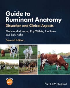
Guide to Ruminant Anatomy: Dissection and Clinical Aspects, 2nd Edition
By Mahmoud Mansour, Ray Wilhite, Joe Rowe and Saly Hafiz
Guide to Ruminant Anatomy: Dissection and Clinical Aspects, 2nd Edition PDF provides a richly illustrated guide tailored to the practical needs of veterinary clinicians. Divided for ease of use into sections representing different parts of the ruminant body, this in-depth introduction uses real dissection images to familiarize readers in detail with the internal and external anatomy of caprine, ovine, and bovine animals. It provides an outstanding demonstration of the relevance of anatomy in clinical settings.
Guide to Ruminant Anatomy readers will also find:
Practical clinical applications discussed by board certified clinicians in each chapter
Line drawings corresponding to dissection images of embalmed specimens
Learning objectives in each section, tying key concepts to clinician development
A companion website featuring laboratory videos demonstrating relevant anatomy
Guide to Ruminant Anatomy is an essential guide for veterinary students studying anatomy of food animals, as well as veterinary practitioners of all kinds looking for an easy-to-use reference on ruminant anatomy.
Table of Contents
Preface xiii
Acknowledgments xv
About the Companion Website xvii
1 The Head, Neck, and Vertebral Column 1
1.1 Skull 2
1.2 Mandible 9
1.3 Paranasal Sinuses 9
1.4 Vertebral Column 14
1.5 Teeth and Age Estimation of Cattle and Small Ruminants (Goats and Sheep) 16
1.5.1 Definitions and Criteria for Estimating the Age of Ruminants 20
1.5.2 Steps for Estimating the Age of Cattle 22
1.6 Joints of the Head 24
1.6.1 Temporomandibular Joint 24
1.6.2 Atlantooccipital Joint 24
1.6.3 Mandibular Symphysis 24
1.6.4 Vertebral Joints 25
1.7 Muscles of the Head 25
1.7.1 Cutaneous Muscles 27
1.7.2 Muscles of Facial Expression 28
1.7.3 Muscles of Mastication 28
1.7.4 Pharyngeal Muscles 28
1.7.4.1 Nomenclature of Pharyngeal Muscles 29
1.7.5 Laryngeal Muscles 29
1.7.6 Hyoid Muscles 29
1.7.7 Lingual Muscles 29
1.7.8 Extraocular Muscles 30
1.8 Blood Vessels, Lymph Nodes, and Nerves of the Head 30
1.8.1 Blood Vessels (Arteries and Veins) 30
1.8.1.1 Arteries of the Head 30
1.8.1.2 Veins of the Head 33
1.8.2 Lymph Nodes of the Head and Neck 33
1.8.3 Nerves of the Head 36
1.8.3.1 Summary of Cranial Nerves and Their Functions 36
1.9 Salivary Glands 40
1.10 The Pharynx 40
1.10.1 Oropharynx 40
1.10.2 Nasopharynx 40
1.10.3 Laryngopharynx 41
1.11 Tongue 41
1.12 The Larynx and Hyoid Apparatus 43
1.12.1 Larynx 43
1.12.2 Hyoid Apparatus 44
1.13 The Eye 45
1.13.1 Superficial Features of the Eye 45
1.13.2 Layers of the Eye 47
1.13.3 Sectioning of the Eyeball 49
1.13.3.1 Chambers of the Eye 49
1.13.4 Drainage Pathway of the Aqueous Humor 49
1.14 Neck Skeleton 49
1.15 Neck Muscles, Nerves, and Vessels 51
1.15.1 Neck Muscles 51
1.15.1.1 Superficial Neck Muscles 54
1.15.1.1.1 Brachiocephalicus Muscle 54
1.15.1.1.2 Omotransversarius Muscle 54
1.15.1.1.3 Trapezius Muscle 55
1.15.1.1.4 Sternocephalicus Muscle 55
1.15.1.1.5 Sternothyroideus and Sternohyoideus Muscles 55
1.15.1.2 Deep Neck Muscles 55
1.15.2 Nerves of the Neck 56
1.15.3 Blood Vessels of the Neck 57
1.16 Nuchal Ligament 57
1.17 Surface Topography (Head and Neck) 58
1.18 Lab ID List for the Head and Neck 62
2 The Thorax 65
2.1 Introduction 66
2.2 Bones of the Thorax 66
2.3 Thoracic Inlet 67
2.4 Basal Border of the Lung and Area for Lung Auscultation 68
2.5 Diaphragmatic Line of Pleural Reflection 68
2.6 Muscles of the Thoracic Wall 69
2.7 Pleura 72
2.7.1 Parietal Pleura 73
2.7.2 Visceral Pleura 73
2.7.3 Connecting Pleura 73
2.7.4 Content of the Pleural Cavity 74
2.7.5 Lung Lobes 74
2.7.6 Mediastinum 74
2.8 Vessels (Arteries and Veins) 75
2.8.1 Blood Circulation: An Overview 75
2.9 Major Veins of the Thorax 76
2.9.1 Cranial Vena Cava 77
2.9.2 Caudal Vena Cava 77
2.9.3 Azygos Veins (Left and Right) 77
2.10 Major Arteries of the Thorax 77
2.10.1 Brachiocephalic Trunk 77
2.10.2 Costocervical Trunk 77
2.10.3 Vertebral Artery 77
2.10.4 Superficial Cervical Artery 78
2.10.5 Internal Thoracic Artery 78
2.11 Lymphatic Structures 78
2.11.1 Thymus 78
2.11.2 Thoracic Duct 78
2.11.3 Mediastinal and Tracheobronchial Lymph Nodes 79
2.12 Nerves (Motor Somatic, Sympathetic, and Parasympathetic) 80
2.12.1 Phrenic Nerve 80
2.12.2 Autonomic Nerves in the Thorax 80
2.12.3 Vagus Nerve 80
2.12.4 Sympathetic Trunk and Sympathetic Ganglia 81
2.13 Heart (Cor) 81
2.13.1 Pericardium 82
2.13.2 External Features of the Heart 82
2.13.3 Interior of the Heart 85
2.13.3.1 Structures of the Right Atrium 85
2.13.3.2 Structures of the Right Ventricle 86
2.13.3.2.1 Right Atrioventricular Valve 86
2.13.3.2.2 Chordae Tendineae, Papillary Muscles, and Cusps of the Right Atrioventricular Valve 87
2.13.3.2.3 Trabecula Septomarginalis 87
2.13.3.2.4 Ossa Cordis 87
2.13.3.2.5 Coronary Arteries 87
2.14 Point of Maximum Intensity or Puncta Maxima 88
2.15 Lab ID List for the Thorax 88
3 The Abdomen 91
3.1 Lumbar Vertebrae 92
3.1.1 Bovine Lumbar Vertebrae 92
3.1.2 Goat and Sheep Lumbar Vertebrae 92
3.2 Ligaments of Lumbar Vertebrae 93
3.2.1 Supraspinous Ligament 93
3.2.2 Interspinous Ligaments 93
3.2.3 Intertransverse Ligaments 93
3.2.4 Yellow Ligaments (Interarcuate or Ligament Flava) 93
3.2.5 Dorsal and Ventral Longitudinal Ligaments 94
3.2.6 Intervertebral Disc 94
3.3 Abdominal Wall 94
3.3.1 Paralumbar Fossa 94
3.3.2 Nerves of the Paralumbar Fossa (Flank Anesthesia) 94
3.3.3 Cutaneus Trunci and Omobrachialis Muscles 95
3.3.4 Tunica Flava Abdominis 98
3.3.5 External Abdominal Oblique Muscle 98
3.3.6 Internal Abdominal Oblique Muscle 101
3.3.7 Transversus Abdominis Muscle 102
3.3.8 Rectus Abdominis Muscle 102
3.3.9 Rectus Sheath 103
3.4 Abdominal Cavity 103
3.4.1 Dissection Plan 105
3.4.2 Peritoneum 105
3.4.3 Omentum 105
3.4.4 Ruminant Stomach 110
3.4.4.1 Reticulum 111
3.4.4.1.1 Cardia 116
3.4.4.3 Rumen 116
3.4.4.3 Omasum 120
3.4.4.4 Abomasum 122
3.5 Intestines 122
3.5.1 Small Intestine 124
3.5.1.1 Duodenum 124
3.5.1.2 Mesoduodenum 125
3.5.1.3 Duodenocolic Fold 125
3.5.1.4 Jejunum 125
3.5.1.5 Ileum 128
3.5.1.6 Ileal Orifice 128
3.5.1.7 Ileocecal Fold 128
3.5.2 Large Intestine 128
3.5.2.1 Cecum and Cecocolic Orifice 129
3.5.2.2 Ascending Colon 129
3.5.2.2.1 Proximal Loop 129
3.5.2.2.2 Spiral Loop 129
3.5.2.2.3 Distal Loop 129
3.6 Other Abdominal Organs 130
3.6.1 Liver 130
3.6.2 Spleen 130
3.6.3 Pancreas 130
3.6.4 Kidney 130
3.7 Vessels 134
3.7.1 Arteries 135
3.7.2 Veins 135
3.7.3 Lymphatics 136
3.8 Palpation of the Live Animal 137
3.9 Lab ID List for the Abdomen 137
4 The Pelvis and Reproductive Organs 139
4.1 Bones of the Pelvis 140
4.1.1 Os Coxae (Pelvic Bone) 140
4.2 Sacrosciatic Ligament 141
4.3 Pelvic Peritoneal Pouches 141
4.4 Urinary Bladder, Ureters, and Ligaments of the Bladder 143
4.5 Male Genitalia 143
4.5.1 Penis 144
4.5.1.1 Root of the Penis and Cavernous Tissue 145
4.5.1.2 Free Part of the Penis and Glans Penis 146