By Gabriel Manso Díaz, Javier López-Sanromán and Renate Weller
A Practical Guide to Equine Radiography PDF is designed to accompany the clinical veterinarian either within a hospital setting or out in the field. The book offers an informative step-by-step guide to obtaining high quality radiographs with a focus on image quality, accuracy, consistency and safety. General principles and equipment are covered before working through the anatomy of the horse with separate chapters devoted to each body region, providing a thorough and detailed picture of the skeletal structure of the horse, making the book an ideal reference for professionals involved with horse health and disease.
Features provided in the book will guide the veterinarian through the stages of taking and interpreting normal radiographs and include:
Clinical indications of radiographic areas of interest in the horse
Equipment required
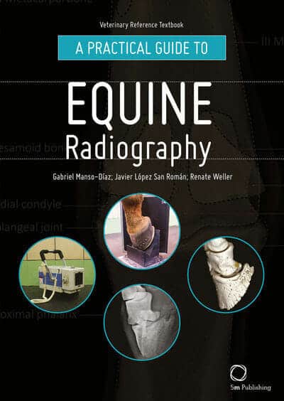
Preparation and setup guides, supported by photographs
Projections focusing on radiographic areas of interest, aided by photographs
x-rays presented with detailed labels, providing a close-up view of skeletal structures
Three dimensional images demonstrating normal anatomy


![Ettinger’s Textbook of Veterinary Internal Medicine 9th Edition [True PDF+Videos]](https://www.vet-ebooks.com/wp-content/uploads/2024/10/ettingers-textbook-of-veterinary-internal-medicine-9th-edition-100x70.jpg "Ettinger’s Textbook of Veterinary Internal Medicine 9th Edition [PDF+Videos]")


