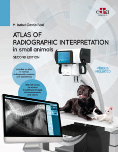
CHAPTER 1: Basic Principles of Radiography
- Fundamentals of radiography
- Definition of X-rays
- X-ray production
- Parts of an X-ray machine
- X-ray interaction with matter
- Image formation
- Radiographic equipment
- The cassette
- Radiographic film
- Image intensifying screens
- Development process
- Negatoscope or X-ray film viewers
- Digital radiography
- Digital X-ray systems
- Native data and image algorithms
- DICOM format
- Software and viewing
- Image archiving and management
- Technical recommendations
- Image quality
- Radiation protection
- International Commission on Radiological Protection
- Legal exposure limits
- Training for exposed workers
- Classification of work areas
- Staff dosimetric control
- Systems and principles for radiation protection
- X-ray interpretation
- Radiographic technique
- Image identification and quality
- Density and contrast
- Image geometry
- Scatter radiation
- Patient positioning and projections
- Technical artifacts and errors
- Radiology reports
- Atlas of Radiographic Positioning and Anatomy
- Abdomen
- Neck
- Thorax
- Appendicular skeleton (thoracic and pelvic limbs)
- Spine
- Head
CHAPTER 2: Abdomen
- Principles of interpretation
- Abdominal wall
- Peritoneal cavity and retroperitoneal space
- Pneumoperitoneum, fluid, peritonitis, carcinomatosis, and masses
- Liver, spleen, and lymph nodes
- Urinary system (kidneys, ureters, bladder)
- Genital system (male and female)
- Stomach and intestines
CHAPTER 3: Neck and Thorax
- Neck structures
- Thoracic wall, ribs, sternum
- Pharynx, larynx, trachea, and esophagus
- Pleural space and mediastinum
- Effusion, pneumothorax, and mediastinal masses
- Heart
- Cardiac diseases (acquired and congenital)
- Lungs and pulmonary diseases
CHAPTER 4: Appendicular Skeleton
- Principles of interpretation
- Bone lesions (osteolysis and osteogenesis)
- Congenital, hereditary, and developmental abnormalities
- Dysplasias, osteochondrosis, fractures, and tumours
- Joint disease and injuries
- Degenerative diseases and immune-mediated disorders
CHAPTER 5: Spine
- Principles of interpretation
- Myelography
- Congenital disorders
- Hemivertebra, block vertebra, and spina bifida
- Instability syndromes and intervertebral disc disease
- Degenerative conditions
- Infections, fractures, and tumours
CHAPTER 6: Head
- Cranial vault
- Hydrocephalus, fractures, infections, and tumours
- Nasal cavity and sinuses
- Mandible, maxilla, and temporomandibular joint
- Teeth and periodontal diseases
- Auditory system
CHAPTER 7: Most Common Diagnostic Errors
- Abdomen: Lack of fat, underexposure, pseudomasses
- Neck and thorax: Skin folds, false cardiomegaly
- Appendicular skeleton: Incorrect positioning, sesamoid bones
- Spine: Overexposure, anatomical variations
- Head: Digital imaging artifacts and overexposure


![Textbook Of Veterinary Diagnostic Radiology 8Th Edition [Pdf+Videos+Quizzes]](https://www.vet-ebooks.com/wp-content/uploads/2019/09/textbook-of-veterinary-diagnostic-radiology-8th-edition.jpg)


![Ettinger’s Textbook of Veterinary Internal Medicine 9th Edition [PDF+Videos] Ettinger’s Textbook of Veterinary Internal Medicine 9th Edition [True PDF+Videos]](https://www.vet-ebooks.com/wp-content/uploads/2024/10/ettingers-textbook-of-veterinary-internal-medicine-9th-edition-100x70.jpg)

![Textbook of Veterinary Diagnostic Radiology 8th Edition [PDF+Videos+Quizzes] Thrall’s Textbook of Veterinary Diagnostic Radiology, 8th edition PDF](https://www.vet-ebooks.com/wp-content/uploads/2019/09/textbook-of-veterinary-diagnostic-radiology-8th-edition-100x70.jpg)


