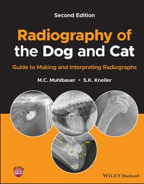
By M. C. Muhlbauer and S. K. Kneller
Radiography of the Dog and Cat: Guide to Making and Interpreting Radiographs, 2nd Edition delivers a thorough update to a celebrated reference manual for all veterinary personnel, student to specialist, involved with canine and feline radiography. The book takes a straightforward approach to the fundamentals of radiography and provides easy-to-follow explanations of key points and concepts. Hundreds of new images have been added covering normal radiographic anatomy and numerous diseases and disorders.
Perfect for veterinary practitioners and students, the second edition of Radiography of the Dog and Cat: Guide to Making and Interpreting Radiographs is also a valuable handbook for veterinary technical staff seeking a one-stop reference for dog and cat radiography.
Features
Features:
- An expanded positioning guide along with images of properly positioned radiographs.
- Numerous examples of radiographic artifacts with explanations of their causes and remedies.
- Detailed explanations of many contrast radiography procedures, including indications, contraindications, and common pitfalls.
- Comprehensive treatments of Musculoskeletal, Thoracic, and Abdominal body parts, including both normal and abnormal radiographic appearances and variations in body types.
Table of Contents
Table of Contents:
-
About the Companion Website
-
Introduction
-
X-Rays
-
Introduction
-
X-ray machine
-
Image receptors
-
Geometry of the x-ray beam
-
X-ray interactions with matter
-
Radiographic density
-
Opacity
-
Radiographic contrast
-
Radiographic detail
-
Technique chart
-
Radiograph storage and distribution
-
Radiation safety
-
Radiographs
-
Introduction – plan for success
-
Positioning guide
-
Artifacts
-
Contrast radiography
-
Procedures in contrast radiography
-
Alimentary tract contrast studies
-
Urogenital contrast studies
-
Cardiovascular contrast studies
-
Neurologic contrast studies
-
Head contrast studies
-
Miscellaneous contrast studies
-
Reading radiographs
-
Thorax
-
Introduction
-
Radiographic views of the thorax
-
Patient factors
-
Thoracic wall
-
Diaphragm
-
Pleura and Pleural Space
-
Mediastinum
-
Esophagus
-
Heart
-
Major vessels
-
Congenital heart disease
-
Acquired heart disease
-
Trachea
-
Lungs
-
Specific lung diseases
-
Differential diagnoses for thorax
-
Abdomen
-
Introduction to abdominal radiography
-
Patient factors
-
Abdominal cavity
-
Liver
-
Spleen
-
Pancreas
-
Gastrointestinal tract
-
Stomach
-
Small intestine
-
Large intestine
-
Urogenital tract
-
Kidneys and ureters
-
Urinary bladder
-
Urethra
-
Male genital system
-
Female genital system
-
Hermaphroditism
-
Adrenal glands
-
Differential diagnoses for abdomen
-
Musculoskeleton
-
Introduction to musculoskeletal radiography
-
Soft tissues
-
Orthopedic anatomic considerations
-
Bone response to disease or injury
-
Bone production
-
Bone loss
-
Fractures
-
Osteomyelitis
-
Osseous neoplasia
-
Benign conditions of bone
-
Congenital and developmental abnormalities
-
Joints
-
Joint diseases
-
Appendicular skeleton
-
Shoulder
-
Elbow
-
Carpus
-
Digits
-
Pelvis
-
Stifle
-
Tarsus
-
Axial skeleton
-
Vertebral column
-
Spinal abnormalities
-
Head and neck
-
Temporomandibular joint (TMJ)
-
Teeth
-
Salivary glands and nasolacrimal duct
-
Pharynx and larynx
-
Differential diagnoses for musculoskeleton
-
Glossary of Radiologic Terms
Index


![Textbook of Veterinary Diagnostic Radiology 8th Edition [PDF+Videos+Quizzes] Thrall’s Textbook of Veterinary Diagnostic Radiology, 8th edition PDF](https://www.vet-ebooks.com/wp-content/uploads/2019/09/textbook-of-veterinary-diagnostic-radiology-8th-edition-235x300.jpg)


![Ettinger’s Textbook of Veterinary Internal Medicine 9th Edition [PDF+Videos] Ettinger’s Textbook of Veterinary Internal Medicine 9th Edition [True PDF+Videos]](https://www.vet-ebooks.com/wp-content/uploads/2024/10/ettingers-textbook-of-veterinary-internal-medicine-9th-edition-100x70.jpg)

![Textbook of Veterinary Diagnostic Radiology 8th Edition [PDF+Videos+Quizzes] Thrall’s Textbook of Veterinary Diagnostic Radiology, 8th edition PDF](https://www.vet-ebooks.com/wp-content/uploads/2019/09/textbook-of-veterinary-diagnostic-radiology-8th-edition-100x70.jpg)


