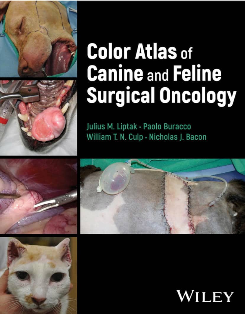
back to top
🔥 Exclusive Lifetime Deal: Unlock Lifetime Updates with a One-Time Payment
Never miss a new book!
Sign Up Now
Home Blog Page 9

Antimicrobial Therapy in Veterinary Medicine 6th Edition

Antimicrobial Therapy in Veterinary Medicine 6th Edition PDF

Antimicrobial Therapy In Veterinary Medicine, 6Th Edition Pdf Download

By Patricia M. Dowling, John F. Prescott and Keith E. Baptiste

Antimicrobial Therapy in Veterinary Medicine 6th Edition has been updated to reflect advances in the field, including new international contributors and a broader global outlook. It includes extensive knowledge of both general principles of mechanisms of antimicrobial drug action including specific classes of antimicrobial agents, as well as chapters dedicated to antimicrobial drug use in a wide range of animal species. As antimicrobial resistance increases as a major global issue in both human and animal health, this book’s renewed focus on antimicrobial stewardship in companion animals, in food animals, and on global aspects keeps it at the forefront of this vital field.

Features
  • Updates to every chapter, reflecting new developments and research, with a complete examination of the issues associated with antimicrobial resistance
  • A comprehensive reference for all aspects of antimicrobial use in veterinary medicine, encompassing theory and practice
  • A global perspective on antimicrobial therapy, with more international content than previous editions
  • A stronger emphasis on antimicrobial stewardship, with practical guidance for prescribing antimicrobial drugs
Table of Contents

Section I: General Principles of Antimicrobial Therapy

  1. Introduction to Antimicrobial Drug Action and Interaction
  2. Antimicrobial Susceptibility Testing
  3. Antimicrobial Resistance
  4. Pharmacokinetics of Antimicrobials
  5. Pharmacodynamics of Antimicrobials
  6. Principles of Antimicrobial Drug Selection and Use

Section II: Classes of Antimicrobial Agents

7. Beta-lactam Antibiotics: Penicillins
8. Beta-lactam Antibiotics: Cephalosporins
9. Other Beta-lactam Antibiotics
10. Peptide Antibiotics
11. Lincosamides, Pleuromutilins, and Streptogramins
12. Macrolides, Azalides, and Ketolides
13. Aminoglycosides and Aminocyclitols
14. Tetracyclines
15. Chloramphenicol and Related Agents
16. Sulfonamides and Combinations
17. Fluoroquinolones
18. Miscellaneous Antimicrobials
19. Antifungal Chemotherapy

Section III: Antimicrobial Stewardship
20. General Concepts in Antimicrobial Stewardship
21. Global Aspects of One Health Antimicrobial Stewardship
22. Antimicrobial Stewardship in Companion Animals
23. Antimicrobial Stewardship in Food-producing Animals
24. Antimicrobial Prophylaxis and Metaphylaxis
25. Regulation of Antimicrobial Use in Animals
26. Antimicrobial Drug Residues in Food

Section IV: Antimicrobial Therapy in Selected Animal Species
27. Horses
28. Dogs and Cats
29. Beef Cattle
30. Dairy Cattle
31. Sheep and Goats
32. New World Camelids
33. Swine
34. Poultry
35. Companion Birds
36. Rabbits, Rodents, and Ferrets
37. Reptiles
38. Zoo and Wildlife Species
39. Aquaculture
40. Honey Bees

Index

Get This Book

This Book is Available For Premium Members Only

Become a Premium Now

Veterinary Surgical Instruments List: Names and Pictures

As a Completion To the Previous Article about Veterinary Equipment and Tools List, In this blog post, we will take a look at some common Veterinary Surgical Equipment List With Names, Uses, and Pictures in veterinary clinics.

Veterinary surgery is an important branch of veterinary medicine, we can’t begin learning veterinary surgery without knowing all the surgical tools you will use in any surgery that’s why we make this Veterinary Surgical Instruments List for all veterinary students to know all Veterinary Surgical Instruments List or veterinarians have come across the issue of needing to purchase surgical instruments for clinics.

Veterinary Books

???? General Surgical Instruments

General veterinary surgical instruments are the basic tools used in all operations. These instruments must be manufactured with high precision and years of experience to ensure the best results. Similarly, the user must be well-trained and experienced to achieve optimal surgical outcomes.

Below are commonly used general instruments in veterinary surgery:

Scissors

Scissors are essential tools used by surgeons to cut and dissect tissues, muscles, organs, and sutures. They allow for the fast and safe removal of obstructive tissues during procedures. Made from balanced stainless steel, surgical scissors are thinner, sharper, and pointed for precision cutting.

Instrument Use Image
Mayo Soft tissue cutting Mayo Scissors
Metzenbaum Soft tissue cutting
Metzenbaum Scissors
Spencer Stitch Suture removal
Spencer Scissors
Standard Suture removal
Standard Scissors
Careless Suture removal
Careless Scissors
Lister Bandage cutting
Lister Scissors
Wire Scissors Cut stainless steel wire
Wire Scissors
Knowles Bandage Scissors Remove bandage and dressing
Knowles Scissors

Forceps

Forceps are essential surgical tools used to hold tissues, separate structures, improve access, and assist in cutting and suturing. They are also used for tweezing and applying pressure during procedures.

Dissecting Forceps

Dissecting forceps are temporary tools used to handle skin, tissues, and organs during surgery. Surgeons also use them to guide needles or manipulate delicate structures.

Type Use Image
Standard plain & toothed Handle soft tissues Standard Forceps
Mosquito Hold small capillaries Mosquito Forceps
Adsons Handle soft tissues Adsons Forceps
Continental Standard Handle skin Continental Forceps
Emmett Handle deep tissues Emmett Forceps
Debakey Handling of viscera Debakey Forceps

Tissue Forceps

Tissue forceps are used for delicate manipulation of tissues without causing trauma. They provide secure grip and precise handling of soft organs and structures during surgical procedures.

Type Use Image
Babcock Handling viscera and soft tissue Babcock Forceps
Allis Handling soft tissue Allis Forceps
Duval Handling viscera and tissue Duval Forceps

Clamps or Hemostats

Hemostats are essential surgical instruments used to occlude blood vessels either completely or partially. They help control bleeding during surgery and are commonly used to block blood flow to internal organs temporarily to protect them during procedures.

Visceral Clamps

Used to occlude visceral organs like the stomach, intestines, or cervix.

Type Use Image
Doyen Mayo Robinson Occlusion of stomach and intestine
Doyen Mayo Robinson Clamp
Parker Kerr Occlusion of cervix
Parker Kerr Clamp

Towel Clamps

Used for securely attaching surgical drapes to the patient’s skin.

Type Use Image
Cross Action Attach drapes to the surgical area
Cross Action Towel Clamp
Backhaus Attach drapes to the surgical area
Backhaus Towel Clamp

Scalpel

The scalpel is a long, thin surgical instrument used to make precise cuts in the skin and underlying tissues. It is an essential tool in dermatology and surgery, enabling incisions, tissue dissections, and various surgical techniques. The term “scalpel” originates from Latin, meaning “small knife.” Scalpels come in a range of sizes and shapes, each suited for specific procedures.

Type Use Image
Scalpel Handle Hold surgical blades
Scalpel Handle
Scalpel Blades Make incision and tissue transection
Scalpel Blades

Retractors

Retractors are used to hold open the incision or wound during surgery, allowing the surgeon better visibility and access to the surgical site. They help retract soft tissues, joints, or organs out of the way, and can be manually held or self-retaining to free the surgeon’s hands.

Manual Retractors

These retractors require manual handling to maintain exposure of tissues and surgical fields.

Type Use Image
Langenbeck Soft tissue retraction
Langenbeck Retractor
Volkman Retraction of tendons and muscles
Volkman Retractor
Hohman Retraction of joints
Hohman Retractor
Czerny Soft tissue retraction
Czerny Retractor

Self-Retaining Retractors

These retractors do not require constant holding and are designed to hold the tissue apart automatically, keeping the surgical field open.

Type Use Image
Gelpi Joint and muscle retraction
Gelpi Retractor
Cone Used in orthopedic surgeries
Cone Retractor
Travers Joint and muscle retraction
Travers Retractor
Gosset Abdominal wall retraction
Gosset Retractor
Balfour Liver retraction
Balfour Retractor
Finochietto Spread ribs
Finochietto Retractor

Needle Holder

A needle holder, also known as a needle driver, is a surgical instrument designed to securely hold suturing needles. It features a comfortable handle for one-handed use and a shaft with a tip that guides the needle through tissues with precision. Some types also incorporate built-in scissors for cutting sutures.

Type Use Image
Gillies Hold needle and cut suture Gillies Needle Holder
Olsen Hegar Hold needle and cut suture Olsen Hegar Needle Holder
Mayo Hegar Hold needle Mayo Hegar Needle Holder
Bruce Clarke Hold needle Bruce Clarke Needle Holder
McPhail Hold needle Mcphail Needle Holder

???? Specific Surgical Instruments

The surgical instruments used by veterinarians during operations must be of the highest quality due to the fragility of organs and bones. Even minor negligence can lead to severe damage. That’s why top-grade, specially designed tools are crucial in veterinary practice.

Orthopedic Surgical Equipment

Orthopedic instruments are used by surgeons to diagnose and treat bone fractures, cut or remove bone, and perform orthopedic procedures. These tools are precision-crafted for efficient and safe handling of skeletal structures.

Instrument Use Image
Chisel Bone shaving
Chisel For Bone Shaving
Gouge Bone shaving
Gouge For Bone Shaving
Osteotome Precise bone cut
Osteotome For Bone Cutting
Periosteal Elevator Raise periosteum before drilling
Periosteal Elevator
Rongeurs Nibble bone pieces
Rongeurs
Mallet To use with chisel
Mallet
Paton Bone Cutting Forceps Cut bone
Paton Forceps
Ruskin Liston Bone Cutting Forceps Cut bone
Ruskin Liston Forceps
Ferguson Bone Holding Forceps Prevent bone movement in surgery
Ferguson Forceps
Kern Bone Holding Forceps Prevent bone movement in surgery
Kern Forceps
Hey Groove Bone Holding Forceps Maintain tip closure
Hey Groove Forceps
Jacob Chuck Insert and remove pins (intramedullary pinning)
Jacob Chuck

Ophthalmic Surgical Instruments

Ophthalmic surgical tools are used for carrying out eye-related surgeries. Both cornea and lens-related surgeries are done with the help of different surgical instruments.

Scissors

Instrument Use Image
Iris scissors To cut iris
Iris Scissors
Castroviejo scissors Cut lens capsule
Castroviejo Scissors
Tenotomy scissors For fine dissection
Tenotomy Scissors

Forceps

Instrument Use Image
Chalazion forceps Stabilize eyelid and protect globe
Chalazion Forceps
Benet cilia forceps For plucking eyelashes
Benet Cilia Forceps
Catford forceps Help in risk-free suturing
Catford Forceps
Capsulorhexis forceps Grasp lens capsule
Capsulorhexis Forceps
Micro corneal tying forceps Tie suture material
Micro Corneal Tying Forceps
Capsule forceps Grabs lens capsule
Capsule Forceps

Hooks

Instrument Use Image
Kirby expressed hook Use for lens removal
Kirby Hook

Speculums

Instrument Use Image
Williams speculum Provide access to eyeball by a retraction
Williams Speculum
Barraquer speculum Provide access to eyeball by a retraction
Barraquer Speculum

Dilators

Instrument Use Image
Nettleship dilator Dilate narrow canals
Nettleship Dilator

Needle Holder

Instrument Use Image
Castroviejo needle holder Hold needle during suturing
Castroviejo Needle Holder

Dental Instruments

Dental instruments are very important for every single dental surgery. They are the most common tools used by vets for the diagnosis and treatment of oral problems—such as removing dental calculus, repairing teeth, and performing extractions. Instruments like scalers, elevators, curettes, and chisels each serve specific functions. Below are commonly used dental surgical instruments:

Instrument Use Image
Dental elevator To separate connection of teeth and bone
Dental Elevator
Extraction forceps Extract tooth
Extraction Forceps
Periosteal elevator To expose bone
Periosteal Elevator
Subgingival curette Remove the unwanted material from the mouth
Subgingival Curette
Dental explorer Expose the hard surface of teeth
Dental Explorer
Supragingival scalers Remove supragingival calculus
Supragingival Scalers
Periodontal probe Measure depth of periodontal pocket
Periodontal Probe
Sharpening stone For sharpening instruments
Sharpening Stone

Teat Instruments

Cows play a crucial role in dairy farming by providing milk, an essential dietary component. However, their teats are prone to damage or infections, which can hinder milk production. Teat surgical instruments are used to treat these issues effectively and maintain udder health. Below are common teat surgical tools:

Instrument Use Image
Dilator Open teat canal
Dilator
Slitter Clear the teat canal by incision from inside to outside
Slitter
Tumor extractor Remove fibrous material from teat canal
Tumor Extractor
Udder infusion canula Administer medicine in teat canal
Udder Infusion Canula
Teat curette Clean inside of teat canal
Teat Curette
Lichty teat knife Open stenotic teats
Lichty Teat Knife
Milking tubes Keep injured teat open
Milking Tubes

Plastic Surgery Instruments

Plastic surgery is a form of body modification that can be approached for a myriad of reasons – whether it is for someone who wants to improve their appearance or for someone who needs to reconstruct parts of their body that have been damaged by injury or disease…

Clamps

Instrument Function Image
Kelly clamp Hold heavy tissues
Kelly Clamp Hold Heavy Tissues
Crile clamp Hold suture ends
Crile Clamp Hold Suture Ends
Rankin clamp Clamp tissues for ligation
Rankin Clamp Clamp Tissues For Ligation
Rochester Pean clamp Clamp larger vessels
Rochester Pean Clamp Clamp Larger Vessels
Moynihan clamp Suturing of vascular tissues
Moynihan Clamp Suturing Of Vascular Tissues
Schmidt clamp Suturing stalk of tissues
Schmidt Clamp Suturing Stalk Of Tissues
Kocher clamp Grasp heavy tissues
Kocher Clamp Grasp Heavy Tissues

Forceps

Instrument Function Image
Dressing forceps Dressing wounds
Dressing Forceps Dressing Wounds
Adson forceps Hold dressing material
Adson Forceps Hold Dressing Material
DeBakey forceps Avoid tissue damage
Debakey Forceps Avoid Tissue Damage
Jansen forceps Remove boney septum
Jansen Forceps Remove Boney Septum
Wilde forceps Stop nasal bleeding
Wilde Forceps Stop Nasal Bleeding

Scissors

Instrument Function Image
Mayo scissors Cut tissue and suture
Mayo Scissors Cut Tissue And Suture
Martin scissors Cut cartilage
Martin Scissors Cut Cartilage
Joseph scissors Cut tissues and sutures
Joseph Scissors Cut Tissues And Sutures
Forman scissors Dissect nasal cartilage
Forman Scissors Dissect Nasal Cartilage
Aufrect scissors Cut delicate tissues
Aufrect Scissors Cut Delicate Tissues
Lister bandage scissors Cut dressing and bandages
Lister Bandage Scissors Cut Dressing And Bandages

Evacuation Instruments

Instrument Function Image
Frazier suction tube Remove debris and fluid from surgical spaces
Frazier Suction Tube Remove Debris And Fluid From Surgical Spaces
Adson suction tube Aspire blood and residues
Adson Suction Tube Aspire Blood And Residues
Baron suction tube Remove fluid and debris
Baron Suction Tube Remove Fluid And Debris

Retractor and Exposure

Instrument Function Image
Army Navy retractor Retract bone and skin
Army Navy Retractor Retract Bone And Skin
Murphy retractor Retract delicate tissues
Murphy Retractor Retract Delicate Tissues
Bear claw retractor Help in a facelift
Bear Claw Retractor Help In A Facelift
Gilles skin hook Retract skin
Gilles Skin Hook Retract Skin
Jansen retractor Spread tissues
Jansen Retractor Spread Tissues

Gastrointestinal Instruments

The digestive system is an important part of the body! It’s responsible for taking in nutrients from your food and turning it into energy. The upper GI tract (esophagus, liver, stomach, gallbladder, spleen, pancreas, diaphragm) and the lower GI tract (small and large bowel, mesentery, appendix, rectum, omentum, anus) each require specialized surgical instruments. Upper‑GI tools are medium‑length; lower‑GI tools are long or extra‑long.

Cholecystectomy Instruments

Instrument Function Image
Collin gallbladder forceps Holding and removal of gallstones
Collin Gallbladder Forceps
Lovelace gallbladder forceps Grab gallstones
Lovelace Gallbladder Forceps
Mixter gallstone forceps Manipulate stone with precision
Mixter Gallstone Forceps
Mayo gallstone scoop Scoop out gallstone
Mayo Gallstone Scoop
Ferguson gallstone scoop Remove stones from gallbladder
Ferguson Gallstone Scoop
Oschner trocar Drain fluids from cavities
Oschner Trocar
Kidney stone forceps Remove stones from bile duct
Kidney Stone Forceps

Liver & Stomach Surgical Instruments

Instrument Function Image
Wishbone retractor Abdominal tissue retraction
Wishbone Retractor
Bookwalter retractor Increase access to surgical site
Bookwalter Retractor
Benson pylorus dilator Grip organs & membranes
Benson Pylorus Dilator
Esophageal dilator Dilate esophagus
Esophageal Dilator
Mayo‑Robson forceps Hold bowel
Mayo‑Robson Forceps
Mayo‑Noble scissors Dissect tissues & skin
Mayo‑Noble Scissors
Scudder intestinal clamp Clamp blood vessels
Scudder Intestinal Clamp

Lower GIT Instruments

Instrument Function Image
Doyen clamp Atraumatic grasping
Doyen Clamp
Dennis clamp Hold tissues
Dennis Clamp
Foss intestinal clamp Clamp bowel
Foss Intestinal Clamp
Fehland intestinal clamp Clamp bleeding site
Fehland Intestinal Clamp
Bainbridge forceps Lock bleeding vessels
Bainbridge Forceps
Dubois scissors Deep dissection
Dubois Scissors
Busch scissors Cut umbilical cords
Busch Scissors
Terminal end stapler Close enterotomies
Terminal End Stapler
Intraluminal stapler Seal tissues in colostomy
Intraluminal Stapler
GI anastomosis stapler Staple transected tube
Gi Anastomosis Stapler

Rectal & Anal Instruments

Instrument Function Image
Fergusen angiotribe clamp Occlude veins and arteries
Fergusen Angiotribe Clamp
Buie pile clamp Grasp hemorrhoids
Buie Pile Clamp
McGivney hemorrhoid ligator Remove hemorrhoids
Mcgivney Hemorrhoid Ligator
Miller rectal scissors Dissect tissues in anal canal
Miller Rectal Scissors
Sims scissors Cut bandages & dead tissues
Sims Scissors
Kelly fistula scissors Cut tissues
Kelly Fistula Scissors
Quimby scissors Cut delicate tissues
Quimby Scissors
Yeoman biopsy forceps Used for diagnosis
Yeoman Biopsy Forceps
Buie fistula probe View anal sac
Buie Fistula Probe
Pratt rectal probe Remove O‑rings
Pratt Rectal Probe
Rosser crypt hook Remove foreign objects
Rosser Crypt Hook
Sawyer retractor Provide access to lower rectum
Sawyer Retractor
Hirschman anoscope Visualize lower rectum
Hirschman Anoscope
Fansler‑Ives anoscope Examine incision area
Fansler‑Ives Anoscope
Hirschman proctoscope Visual inspection of anal area
Hirschman Proctoscope
Rigid sigmoidoscope Examine inside of anus
Rigid Sigmoidoscope
Chelsea‑Eaton anal speculum View rectum
Chelsea‑Eaton Anal Speculum
Pratt rectal speculum Dilate anal area
Pratt Rectal Speculum

Cardiothoracic And Vascular Surgical Instruments

In the field of cardiothoracic surgery, common procedures include coronary artery bypass grafting and aortic valve replacement. Although approaches and incisions differ, surgical training and expertise are key to outcomes. Advances in medical equipment have given surgeons specialized tools to remove vessels, repair valves, and perform bypasses with greater precision and safety.

Clamps & Related Instruments

Instrument Function Image
Rochester‑Pean clamp Clamp larger vessels
Rochester‑Pean Clamp
Kantrowitz clamp Grasp tissues
Kantrowitz Clamp
Finochietto clamp Grasp tissues & blood vessels
Finochietto Clamp
Bulldog applicator Apply bulldog clamp
Bulldog Applicator
Sweet clip applier Ligate tubular sutures
Sweet Clip Applier
Gluck rib shears Cut ribs
Gluck Rib Shears
Doyen rib stripper Strip periosteum from ribs
Doyen Rib Stripper

Probes & Dilators

Instrument Function Image
Garrett vascular dilators Perform vessel dilation Garrett Vascular Dilators

Measuring Instruments

Instrument Function Image
Tessier caliper Measure anatomical structures Tessier Caliper

Installation & Evacuation Instruments

Instrument Function Image
Wolf suction Remove blood from surgical site
Wolf Suction
Poole suction tube Remove large volumes of fluid
Poole Suction Tube

Retraction & Exposure

Instrument Function Image
Allison retractor Retract lungs
Allison Retractor
Malleable ribbon retractor Retract during orbital dissection
Malleable Ribbon Retractor
Leaflet retractor Used in cardiac surgery
Leaflet Retractor

Special Instruments

Instrument Function Image
Vessel punch Create an opening in vessels
Vessel Punch
McIntosh suture holder Separate sutures
Mcintosh Suture Holder
Rummel tourniquet Occlude blood vessels
Rummel Tourniquet

Endoscopic Instruments

Endoscopy is used in animals to examine internal organs via a camera, either diagnostically (viewing the digestive tract through mouth or anus) or therapeutically (removing foreign objects, repairing abnormalities). Under general anesthesia, veterinarians can visualize the heart, lungs, stomach, intestines, and more. Endoscopy improves recovery by avoiding large incisions.

Essential Endoscopic Instruments

Instrument Function Image
Endoscopic trocar Small‑puncture port
Endoscopic Trocar
Veress needle Establish pneumoperitoneum
Veress Needle
“S” retractors Retract abdominal walls
S Retractors
Thread sleeve Reinforce suture connection
Thread Sleeve
Balloon dissector Separate extraperitoneal tissue
Balloon Dissector
Insufflation tubing Insufflate abdominal cavity
Insufflation Tubing

Viewing of Working Space

Instrument Function Image
Fiber‑optic light cable Illuminate surgical field
Fiber‑Optic Light Cable
Telescope Magnified internal view
Telescope
Camera head Capture endoscopic images
Camera Head
Bipolar cord Connect bipolar instruments
Bipolar Cord
Monopolar cord Connect monopolar instruments
Monopolar Cord

Special Instruments

Instrument Function Image
Chitwood DeBakey clamp Clamp lung tissues
Chitwood Debakey Clamp
Dennis clamp Hold tissues
Dennis Clamp
Chitwood suture cutter Cut sutures
Chitwood Suture Cutter

Other Equipment

Below are additional miscellaneous instruments commonly used in various veterinary surgical procedures.

Instrument Use Image
Cusco Vaginal Speculum Exposure of vaginal tissue
Cusco Vaginal Speculum
Rampley Sponge Holding Forceps Hold swabs and sponges
Rampley Sponge Holding Forceps
Hartman Crocodile Forceps Nasal and oral use
Hartman Crocodile Forceps
For More Vet Surgical Instruments Download This PDF Guide:

Conclusion

There are many reasons why surgical instruments are needed in surgeries. First, they are used to cut and pierce the skin. Second, they are used to perform the operation. Third, they are used to remove foreign objects. Fourth, they are used to keep the patient from bleeding. So that’s why we need surgical instruments for surgeries. We hope you enjoyed our article about surgical instruments. You may have never thought about it, but there are a lot of different instruments that are used in surgeries. Understanding the different instruments and what they are used for will help you appreciate the skill and expertise of veterinary surgeons. Since there are so many different instruments used in surgeries, it can be difficult to keep track of them all but we have mentioned the most important ones.

Tip

Do You Want To Increase Your Veterinary Knowledge and Practical Skills?

You Can Now Browse and Download +3000 Veterinary Books Online In All Veterinary Fields.

Register Now

Behavior Problems of the Dog and Cat 4th Edition

Behavior Problems Of The Dog And Cat 4Th Edition Pdf Download

By Gary Landsberg, Wayne Hunthausen and Lowell Ackerman

Behavior Problems of the Dog and Cat 4th Edition retains the highly practical approach that has proved so successful in previous editions, offering diagnostic guidelines, preventive advice, treatment guidelines and charts, case examples, client forms and handouts, and product and resource suggestions, along with details on the use of drugs and natural supplements to help optimize the behavior services offered in practice.

Features

Behavior Problems of the Dog and Cat Features:

  • Step-by-step guidelines describe how to collect a patient history, perform a thorough physical examination, conduct diagnostic testing, formulate differential diagnoses, select treatment, and monitor the patients’ responses.
  • Background information describes how dog and cat behavior problems arise and how they can be prevented.
  • Coverage of behavior modification techniques provides you with a clear understanding of suggested treatment as well as the use of drugs, products, pheromones, surgery, diet, and alternative therapies.
  • Content on behavioral genetics explores this rapidly growing and advancing field and includes new therapeutic approaches for cognitive decline.
  • Case studies illustrate real-life clinical situations.
  • Easy-access treatment tables provide at-a-glance solutions to common behavior problems.
  • Useful appendices include treatment protocols as well as the drug information and dosages that make effective prescribing easy.
  • NEW! Many hot new topics are covered, including fear, anxiety, and stress and their effects on health and behavior, as well as pain and behavior, the psychobiological approach to veterinary behavior assessment, and pets and the family dynamic.
  • NEW! Updated chapter content is extensively augmented or completely rewritten by new authors, making this more than just a new edition – it’s a new book!
  • NEW! eBook version is included with print purchase which allows access to all of the text, figures, and references, with the ability to search, customize content, make notes and highlights, and have content read aloud. Online access also includes handouts and forms, drug dosing, and a comprehensive directory of resources.
Table of Contents
  1. Behavioral medicine and the general practitioner
  2. Developmental, social, and communicative behavior
  3. Pet selection and the genetics of behavior
  4. Pets and the family dynamic
  5. Prevention: the best medicine
  6. All body systems affect behavior
  7. Physiologic stress and its effect on health and welfare
  8. The effects of aging on behavior in senior pets
  9. Approach to the diagnosis and treatment
  10. Behavioral treatment techniques, behavior modification, and learning theory
  11. Pharmacologic intervention in behavioral therapy
  12. Treatment – integrative medicine
  13. Feeding and diet-related problems
  14. Fears, phobias, and anxiety disorders
  15. Noise aversion
  16. Reducing fear, anxiety, and stress in veterinary clinics
  17. Separation-related disorders
  18. Abnormal repetitive behaviors: stereotypies and compulsive disorders
  19. Unruly and destructive behaviors – canine
  20. Unruly and destructive behaviors – feline
  21. House soiling – Canine
  22. House soiling – feline
  23. Canine aggression
  24. Feline aggression
  25. The psychobiological approach to problem behavior assessment
  26. Appendix A – Behavior resources
    Appendix B – Medication dosages
    Appendix C – Resources available from book website
    Appendix D – This appendix contains forms and handouts that are available online in printable format.
  27. Index

Get This Book

This Book is Available For Premium Members Only

Become a Premium Now

Bill’s Clinical Pharmacology and Therapeutics for Veterinary Technicians 5th Edition

Bill's Clinical Pharmacology and Therapeutics for Veterinary Technicians, 5th Edition

Bill's Clinical Pharmacology And Therapeutics For Veterinary Technicians 5Th Edition Pdf Download

By Melinda Anderson

Bill’s Clinical Pharmacology and Therapeutics for Veterinary Technicians helps you understand the principles and practice of pharmacology in treating different animal species. The text explains how drugs work, how they are administered, and how to calculate drug dosages. Covering drugs by body system, the book also explains possible abuses, mistakes, and how things can go wrong when therapeutic protocols are not properly implemented. From veterinary pharmacist and educator Melinda “Mindy” Anderson, Bill’s Clinical Pharmacology and Therapeutics for Veterinary Technicians, 5th Edition helps you provide better care for your patients and better education for their owners.

Features
  • Content on basic pharmacology includes topics such as safety, terminology, pharmacy procedures, drug handling (including the latest OSHA requirements), dosage calculations, and pharmacokinetics.
  • Chapter outlines, learning objectives, and key terms are provided at the beginning of each chapter, introducing you to the complex principles of pharmacology and guiding your study.
  • Clear explanations of drugs help you understand the “how” and “why” behind drugs, their actions, their mechanisms, and adverse effects.
  • Myths and Misconceptions, Ask Dr. Bill, and You Need to Know boxes spotlight key issues, concepts, and skills.
  • Clinical applications link pharmacologic concepts to real-life situations.
  • Review questions and critical thinking questions are included at the end of every chapter to help readers test their comprehension.
  • Test questions and online quizzes enable self-assessment and help you prepare for classroom and certification exams.
Table of Contents
  1. Veterinary Pharmacology and the Veterinary Technician
  2. Pharmacy Procedures and Dosage Calculations
  3. Pharmacokinetics and Pharmacodynamics: The Principles of How Drugs Work
  4. Drugs Affecting the Gastrointestinal Tract
  5. Drugs Affecting the Cardiovascular System
  6. Drugs Affecting the Respiratory System
  7. Drugs Affecting the Endocrine System
  8. Drugs Affecting the Nervous System: Analgesics, Tranquilizers, Sedatives, and Anesthetics
  9. Drugs Affecting the Nervous System: Anticonvulsants and Behavior-Modifying Drugs
    Antimicrobials
  10. Disinfectants and Antiseptics
  11. Antiparasitics
  12. Antiinflammatory Drugs

Answers to Self Assessment

Glossary

Index

Get This Book

This Book is Available For Premium Members Only

Become a Premium Now

Nutrition and Disease Management for Veterinary Technicians and Nurses 3rd Edition

Nutrition and Disease Management for Veterinary Technicians and Nurses 3rd Edition

Nutrition And Disease Management For Veterinary Technicians And Nurses 3Rd Edition Pdf Download

By Ann Wortinger and Kara M. Burns

Nutrition and Disease Management for Veterinary Technicians and Nurses 3rd Edition offers an accessible, up-to-date introduction to the key concepts and elements of veterinary nutrition. With detailed coverage of the fundamentals of veterinary nutrition in addition to the nutritional management for many common small animal diseases, the third edition incorporates all the essentials of veterinary nutrition and dietary management for veterinary patients of any age or health status.

Nutrition and Disease Management for Veterinary Technicians and Nurses is ideal for all veterinary technician students, working veterinary technicians, nurses, and students working towards specialty certification in veterinary nutrition.

Features
Readers of the third edition of Nutrition and Disease Management for Veterinary Technicians and Nurses will also find:

  • Fourteen new chapters, plus additional species in some chapters
  • An added section on prebiotics, probiotics, and synbiotics
  • End-of-chapter summaries with multiple-choice questions and case reviews to facilitate use in the classroom
Table of Contents

Section I The Basics of Nutrition 

1 Nutrients and Energy

2 Water

3 Carbohydrates

4 Fats

5 Protein and Amino Acids

6 Vitamins

7 Minerals

8 Digestion and Absorption

9 Energy Balance

10 Gastrointestinal Microbiome

11 Prebiotics, Probiotics and Synbiotics

12 Nutrition Calculations

Section II Nutritional Requirements of Dogs and Cats

13 History and Regulation of Pet Foods 91

14 Pet Food Labels 97

15 Nutrient Content of Pet Foods

16 Types of Pet Foods

17 Raw Food Diets

18 Grain-Free and Boutique Diets 

19 Additives and Pet Food Preservatives

20 Homemade Diets

21 Resources for Alternative Diets

Section III Feeding Management for Dogs and Cats 

22 Feeding Regimens for Dogs and Cats

23 Nutritional Assessment

24 Pregnancy and Lactation in Dogs

25 Pregnancy and Lactation in Cats

26 Neonatal Puppies and Kittens

27 Growth in Dogs

28 Growth in Cats

29 Adult Maintenance in Dogs

30 Adult Maintenance in Cats

31 Feeding the Healthy Geriatric Dog and Cat

32 Performance and Dogs

33 Feeding Requirements of Cats

34 Nutrition Myths

35 Cost of Feeding

Section IV Nutritional Disease Management for Dogs and Cats 

36 Nutritional Management of Gastrointestinal Disorders

37 Critical Care Nutrition

38 Assisted Feeding in Dogs and cats

39 Liver Disease

40 Dermatology

41 Fatty Acids in Disease Management

42 Endocrinology

43 Cancer Nutrition

44 Refeeding Syndrome

45 Cardiac Disease

46 Musculoskeletal

47 Weight Management

48 Cachexia

49 Dental Health

50 Nutritional Management of Pancreatitis

51 Nutrition in Pancreatic Insufficiency

52 Nutritional Management of Hyperlipidemia in Dogs and Cats

53 Brain Aging/Cognitive Dysfunction

54 Kidney Disease

55 Nutritional Management of Digestive Disease in Brachycephalic Dogs

56 Feline Lower Urinary Tract Disease

Section V Feeding Management for other Companion Animals 

57 Avian

58 Small Pet Mammals and Reptiles

59 Equine

Index

Get This Book

This Book is Available For Premium Members Only

Become a Premium Now

Animal Restraint for Veterinary Professionals 3rd Edition

Animal Restraint for Veterinary Professionals 3rd Edition

Animal Restraint For Veterinary Professionals 3Rd Edition Pdf Download

By C. C. Sheldon, Teresa Sonsthagen and James Topel

Animal Restraint for Veterinary Professionals 3rd Edition includes photos showing the exact steps needed to achieve safe restraint of animals seen in the veterinary clinic and visited on farm calls. Separate chapters are devoted to each domestic animal species, including cats, dogs, cattle, horses, sheep, goats, pigs, rabbits, rodents, ferrets, and birds. This edition adds descriptions of new restraint techniques for cats, dogs, and other species. Written by expert veterinary technician instructors C.C. Sheldon, and James A. Topel, this practical guide also includes a chapter on knot tying!

Features
  • Step-by-step guidelines make it easy to understand safe, commonly used restraint procedures, and include alternative restraint techniques for several different species.
  • More than 500 full-color images and a clear, non-technical writing style depict and simplify animal restraint concepts.
  • Comprehensive coverage includes normal and abnormal animal behavior characteristics, precautions, special handling, restraint devices, and descriptions of special restraint techniques for medical procedures such as venipuncture.
  • Restraint Principles chapter summarizes the considerations for proper restraint, animal safety, circumstances for restraint, the effect of restraint on animals, and complications.
  • Knot Tying chapter presents basic types of knots that may be used to safely restrain larger animals.
  • NEW! Additional restraint techniques are included for several species.
  • NEW! Restraint of Cats chapter is updated to include guidelines and photos for the removal of cats from a carrier, examination in a carrier, and examination in a towel on the handler’s lap.
  • NEW! Restraint of Dogs chapter is updated to include canine head control using a towel, along with safe techniques for administering intramuscular injections to aggressive dogs including the use of a door as a squeeze area and hip catch.
  • NEW! Learning objectives, chapter outlines, and key terms are added to the beginning of each chapter.
Table of Contents
  1. Restraint Principles
  2. Knot Tying
  3. Restraint of Cats
  4. Restraint of Dogs
  5. Restraint of Cattle
  6. Restraint of Horses
  7. Restraint of Sheep
  8. Restraint of Goats
  9. Restraint of Swine
  10. Restraint of Rodents, Rabbits and Ferrets
  11. Restraint of Birds
  12. Appendix 1: Gender Names
  13. Appendix 2: Physiologic Data
  14. Glossary

Get This Book

This Book is Available For Premium Members Only

Become a Premium Now

Large Animal Clinical Procedures for Veterinary Technicians 5th Edition

Large Animal Clinical Procedures for Veterinary Technicians, 5th Edition

Large Animal Clinical Procedures For Veterinary Technicians 5Th Edition Pdf Download

By Kristin J. Holtgrew-Bohling

Get the big picture on the vet tech’s role and responsibilities in large animal care! A practical, comprehensive guide, Large Animal Clinical Procedures for Veterinary Technicians, 5th Edition describes how to set up, assist, and follow through on medical procedures and treatment regimens for domestic livestock. The book begins with an overview of livestock handling, reproduction, and nutrition, along with the skills required in hospital management. Following are separate sections on horses, cattle, sheep and goats, llamas and alpacas, swine, and poultry, with each section including chapters examining husbandry, clinical and surgical procedures, and common diseases. Written by expert clinician and vet tech educator Kristin Holtgrew-Bohling, this text provides an ideal study tool in preparing for the VTNE® and for everyday practice.

Features
  • Comprehensive large animal coverage is specifically tailored to the needs of veterinary technician students.
  • Coverage of the essential large animal-related tasks in the CVTEA Manual of Accreditation for Veterinary Technology Programs prepares you to pass the Veterinary Technology National Exam (VTNE®).
  • Step-by-step procedures explain how and why a clinical procedure is performed, and the roles that you fill in preparing for, assisting in, and following up the procedure.
  • “How-to” chapters within each species section examine husbandry, clinical procedures, surgical procedures, and common diseases — so veterinary technicians, acting under instructions of veterinarians, can plan and follow through on procedures and treatment regimens for large animals.
  • Evolve website includes quizzes, images, and reference materials to reinforce understanding.
  • Full-color photographs and line drawings show step-by-step procedures in areas such as restraint, bandaging, physical examination techniques, and diagnostic procedures.
  • Coverage of husbandry and breeds demonstrates how day-to-day housing and feeding affect the care of many large animal diseases, and also helps you provide quality client education.
  • Livestock Industry section provides an overview of safety and handling, reproduction, and nutrition, so you can better understand the practices, procedures, and decisions in large animal veterinary medicine.
  • Learning features enhance critical thinking and decision making with case studies, clinical applications, key terms, chapter outlines, learning objectives, and Technician Notes.
Table of Contents

PART 1: The Livestock Industry
1. The Importance of Livestock
2. Livestock Safety and Handling
3. Livestock Reproduction
4. Livestock Nutrition
PART 2: Large Animal Hospital Management
5. Daily Hospital Responsibilities
6. Admissions, Medical Records, and Physical Examinations
7. Diagnostic Imaging for Large Animals
PART 3: Horses in Veterinary Practice
8. Equine Husbandry
9. Equine Clinical Procedures
10. Equine Surgical Procedures
11. Common Equine Disease
PART 4: Cattle in Veterinary Practice
12. Bovine Husbandry
13. Bovine Clinical Procedures
14. Bovine Surgical Procedures
15. Common Bovine Diseases
PART 5: Sheep and Goats in Veterinary Practice
16. Ovine and Caprine Husbandry
17. Ovine and Caprine Clinical Procedures
18. Ovine and Caprine Surgical Procedures
19. Common Ovine and Caprine Diseases
PART 6: Llamas and Alpacas in Veterinary Practice
20. Camelid Husbandry
21. Camelid Clinical Procedures
22. Camelid Surgical Procedures
23. Common Camelid Diseases
PART 7: Swine in Veterinary Practice
24. Porcine Husbandry
25. Porcine Clinical Procedures
26. Porcine Surgical Procedures
27. Common Porcine Diseases
PART 8: Poultry in Veterinary Practice
28. Poultry Husbandry
29. Poultry Clinical Procedures
30. Poultry Surgical Procedures
31. Common Poultry Diseases
Glossary

Get This Book

This Book is Available For Premium Members Only

Become a Premium Now

Atlas of Radiographic Interpretation in Small Animals, 2nd Edition

Atlas of Radiographic Interpretation in Small Animals, 2nd Edition PDF

Atlas Of Radiographic Interpretation In Small Animals, 2Nd Edition Pdf Download

By Isabel M. García Real

Atlas of Radiographic Interpretation in Small Animals, 2nd Edition extends its initial aim of assisting general veterinary practitioners in radiographic interpretation and pathological diagnosis by adding to the content of the first edition a chapter on the principles of radiology and a complete atlas of radiographic positioning and anatomy. It includes 400 new images and updates previous graphic material content to further increase its quality. In addition, the book is complemented by new multimedia material, which can be accessed through QR codes located throughout the text. In this way, the reader can access different normal radiographic anatomy diagrams, both with and without the anatomical details identified.

Table of Contents

CHAPTER 1: Basic Principles of Radiography

  • Fundamentals of radiography
  • Definition of X-rays
  • X-ray production
  • Parts of an X-ray machine
  • X-ray interaction with matter
  • Image formation
  • Radiographic equipment
    • The cassette
    • Radiographic film
    • Image intensifying screens
    • Development process
    • Negatoscope or X-ray film viewers
  • Digital radiography
    • Digital X-ray systems
    • Native data and image algorithms
    • DICOM format
    • Software and viewing
    • Image archiving and management
    • Technical recommendations
    • Image quality
  • Radiation protection
    • International Commission on Radiological Protection
    • Legal exposure limits
    • Training for exposed workers
    • Classification of work areas
    • Staff dosimetric control
    • Systems and principles for radiation protection
  • X-ray interpretation
    • Radiographic technique
    • Image identification and quality
    • Density and contrast
    • Image geometry
    • Scatter radiation
    • Patient positioning and projections
    • Technical artifacts and errors
    • Radiology reports
  • Atlas of Radiographic Positioning and Anatomy
    • Abdomen
    • Neck
    • Thorax
    • Appendicular skeleton (thoracic and pelvic limbs)
    • Spine
    • Head

CHAPTER 2: Abdomen

  • Principles of interpretation
  • Abdominal wall
  • Peritoneal cavity and retroperitoneal space
    • Pneumoperitoneum, fluid, peritonitis, carcinomatosis, and masses
  • Liver, spleen, and lymph nodes
  • Urinary system (kidneys, ureters, bladder)
  • Genital system (male and female)
  • Stomach and intestines

CHAPTER 3: Neck and Thorax

  • Neck structures
  • Thoracic wall, ribs, sternum
  • Pharynx, larynx, trachea, and esophagus
  • Pleural space and mediastinum
    • Effusion, pneumothorax, and mediastinal masses
  • Heart
    • Cardiac diseases (acquired and congenital)
  • Lungs and pulmonary diseases

CHAPTER 4: Appendicular Skeleton

  • Principles of interpretation
  • Bone lesions (osteolysis and osteogenesis)
  • Congenital, hereditary, and developmental abnormalities
    • Dysplasias, osteochondrosis, fractures, and tumours
  • Joint disease and injuries
    • Degenerative diseases and immune-mediated disorders

CHAPTER 5: Spine

  • Principles of interpretation
  • Myelography
  • Congenital disorders
    • Hemivertebra, block vertebra, and spina bifida
  • Instability syndromes and intervertebral disc disease
  • Degenerative conditions
  • Infections, fractures, and tumours

CHAPTER 6: Head

  • Cranial vault
    • Hydrocephalus, fractures, infections, and tumours
  • Nasal cavity and sinuses
  • Mandible, maxilla, and temporomandibular joint
  • Teeth and periodontal diseases
  • Auditory system

CHAPTER 7: Most Common Diagnostic Errors

  • Abdomen: Lack of fat, underexposure, pseudomasses
  • Neck and thorax: Skin folds, false cardiomegaly
  • Appendicular skeleton: Incorrect positioning, sesamoid bones
  • Spine: Overexposure, anatomical variations
  • Head: Digital imaging artifacts and overexposure

Get This Book

This Book is Available For Premium Members Only

Become a Premium Now

Anatomy and Histology of the Domestic Chicken

Anatomy and Histology of the Domestic Chicken PDF

Anatomy And Histology Of The Domestic Chicken
By Josep Rutllant and Wael Khamas

Anatomy and Histology of the Domestic Chicken is a state-of-the-art atlas of avian anatomy that provides a complete collection of both original gross anatomy and histology photographs and texts of all body systems of the birds based on the domestic chicken to depict anatomic features. Using cutting-edge technology to create visualizations of anatomic structures, this exhaustive reference includes both gross anatomical structures/organs and their histological details next to each other. This approach enables readers to understand the macro- and micro-pictures of each organ/structure under study.

Features

Book Features:

  • External features of the body, including regions, features, ornaments, shape, feathers, skin, and the uropygial gland
  • Musculoskeletal characteristics including cartilage and bone formation and classification, as well as flight and ambulatory muscles
  • Digestive system, including the beak, esophagus, crop, proventriculus, ventriculus, intestines, and accessory glands
  • Respiratory system, including external nares, nasal cavity, trachea, upper larynx, syrinx, lungs, and air sacs
  • Urinary system, including kidneys and the ureter, cloaca-urodeum, and genital system, covering differences between males and females
  • Endocrine system, including pituitary, pineal, adrenal, pancreas, thyroid, and parathyroid glands
  • Nervous system with central and peripheral divisions and sense organs including eye and ear
  • Lymphatic system, with descriptions of the primary and secondary lymphatic organs
  • Egg anatomy and development of the chick embryo
  • Applied anatomical concepts important for clinical maneuvers and necropsy
Table of Contents
  • External Features of Chicken
    • Skin (Epidermis, Dermis, Hypodermis)
    • Structures associated with the Skin (Patagium, Operculum, Ornaments)
    • Feathers (Development, Description, Types, Parts, Molting)
    • Lamellar Corpuscles
    • Debeaking
    • Eyelids, Vent, Uropygial Gland
    • Brood Pad, Bursa Sterni
    • Legs (Podotheca, Scutes, Digits, Claws, Spur, Pads)
    • Thermoregulation
  • Skeletal System
    • Introduction to Skeletal System
    • Cartilage and Bone Embryonic Origin
    • Cartilage Cell Types (Chondroprogenitor, Chondroblast, Chondrocyte, Chondroclast)
    • Types of Cartilages (Hyaline, Fibrocartilage, Elastic)
    • Cartilage Development
    • Calcium Homeostasis
    • Bone Cell Types (Mesenchymal, Pericyte, Osteogenic, Osteoblast, Osteocyte, Osteoclast)
    • Ossification (Intramembranous, Endochondral)
    • Axial Skeleton (Skull, Vertebrae, Notarium, Ribs, Sternum)
    • Sesamoid and Pneumatic Bones
    • Pectoral and Pelvic Girdle
    • Pectoral Limb (Wing)
    • Pelvic Limb
    • Arthrosis (Joints)
  • Muscular System
    • Introduction to Muscular System
    • Muscle Histology (Smooth, Cardiac, Skeletal)
    • Muscle Nomenclature
    • Types of Skeletal Muscle Fibers
    • Muscle Cells (Myocyte, Satellite)
    • Skeletal Muscle Blood Supply and Innervation
    • Muscle Spindle
    • Tendon Histology and Modifications
    • General Description of Skeletal Muscle
    • Cutaneous Muscles
    • Flight Muscles (Pectoral, Wing Muscles)
    • Muscles of the Pelvic Region (Thigh, Leg)
    • Abdominal Muscles
  • Digestive System
    • Introduction to Digestive System
    • Oral Cavity and Pharynx (Beak, Tongue, Salivary Glands, Taste Buds, Pharyngoesophageal Junction)
    • Hyoid Apparatus
    • Esophagus
    • Stomach (Proventriculus, Intermediate Zone, Gizzard)
    • Small Intestine (Duodenum, Jejunum, Ileum)
    • Large Intestine (Rectum, Ceca)
    • Cloaca, Vent
    • Liver (Hepatic Portal System and Blood Supply)
    • Pancreas
  • Respiratory System
    • Introduction to Respiratory System
    • Nostrils and Nasal Cavity
    • Larynx
    • Trachea
    • Syrinx
    • Lungs
    • Direction of Airflow
    • Blood-Gas Barrier
    • Air Sacs
  • Urinary System
    • Introduction to Urinary System (Composition, Function, Development)
    • Kidney (Anatomy, Histology)
    • Ureter, Urodeum
    • Blood Supply, Innervation
    • Urine
  • Reproductive System
    • Male Reproductive System (Testes, Epididymis, Deferent Duct, Phallus, Spermatogenesis, Spermatozoa)
    • Female Reproductive System (Ovary, Oviduct, Cloaca)
    • Formation of Chicken’s Egg (Yolk, Albumen, Shell Membranes, Testa)
  • Endocrine System
    • Introduction to Endocrine System
    • Physiological Balance (Homeostasis)
    • Major Endocrine Glands (Hypothalamus, Pituitary, Adrenal, Thyroid, Parathyroid, Ultimobranchial Bodies, Carotid Body, Pineal Gland, Islets of Langerhans, Gonads, Digestive Enterochromaffin Cells)
  • Sense Organs
    • The Eye (Eyeball, Lens, Eye Chambers, Vision)
    • The Ear (External, Middle, Inner, Hearing)
  • Cardiovascular System
    • Introduction to Cardiovascular System
    • Heart (Topography, Pericardial Sac, Surface Anatomy, Chambers, Valves, Conducting System, Arterial Supply, Venous Drainage)
    • General Arterial System (Pulmonary Trunk, Systemic Arteries)
    • Venous System (Pulmonary Veins, Systemic Veins)
    • Blood Collection Sites
    • Hepatic Portal System
    • Renal Portal System
    • Carotid Body/Sinus
  • Lymphatic System
    • Introduction to Lymphatic System
    • Cells of the Lymphatic System (Lymphocytes, Monocytes, Macrophages, Reticular Cells)
    • Primary and Secondary Lymphatic Organs (Bursa of Fabricius, Thymus, Spleen, Cecal Tonsils, Gut-Associated Lymphoid Tissue)
    • Maternal Transfer of Immunity
    • Lymphatic Vessels
  • Nervous System
    • Introduction to Nervous System
    • Neurons (Types, Synapse)
    • Ganglion (Sensory, Autonomic)
    • Glial Cells (Microglial, Macroglial)
    • Peripheral Nervous System Cells (Schwann, Satellite)
    • Meninges
    • Brain Ventricles
    • Cerebrospinal Fluid (CSF)
    • Blood Brain Barrier
    • Cerebrum (Telencephalon, Diencephalon, Mesencephalon, Metencephalon, Myelencephalon)
    • Reticular Formation
    • Spinal Cord
    • Autonomic Nervous System (Sympathetic, Parasympathetic)
  • Applied Chicken Anatomy
    • Examining from a Distance (Behavioral Deviations, Abnormal Mentation, Posture)
    • Physical Examination (Feathers, Head, Wattles, Eyes, Ears, Thorax, Cloaca, Legs)
    • Specialized Clinical Procedures (Identification Placement, Culture Techniques, Blood Collection)
  • Chicken Necropsy
    • Introduction to Necropsy
    • Euthanasia and Carcass Disposal
    • Chicken Disposal
    • Facilities and Materials Needed (Personal Protective Equipment, Histopathological Examination, Collection of Microbiological Samples, Molecular Diagnosis, Toxicological Investigation)
    • Whole Carcass Conservation
    • Necropsy Protocol (External and Internal Examination)
  • The Egg Anatomy
    • Introduction to Egg Anatomy
    • The Germinal Disc
    • The Yolk
    • The Albumen
    • The Eggshell (Shell Membranes, Testa, Cuticle)
  • Fertilization and Chick Embryo Development
    • Introduction to Fertilization and Development
    • Avian Fertilization (Sperm Deposition, Storage, Transport, Ovulation, Fertilization)
    • Chick Embryo Development
  • Index

Get This Book

This Book is Available For Premium Members Only

Become a Premium Now

The Immunology of the Dog

The Immunology of the Dog Book

The Immunology Of The Dog Pdf Download

By Rosina Lillywhite and Marie Rippingale

The Immunology of the Dog Book is the first readily available, single-source, comprehensive review of the canine immune system. The book discusses all immunologic aspects of diseases of the dog and relates recent basic science to important clinical issues. Sections cover the evolution and domestication of the dog, provide detailed information on immunity to infectious diseases, canine allergic diseases, the top canine autoimmune diseases, and canine immunodeficiencies. The Immunology of the Dog offers professionals in veterinary research and practice complete information on all aspects of canine immunology in one essential resource.

  • Introduces the first comprehensive review of canine immunology
  • Links recent basic science to important clinical issues in dogs
  • Provides detailed and current information in one resource
Table of Contents
  1. The Evolution and Domestication of the Dog
  2. Ontogeny, Reproduction, and Lactation
  3. Canine Innate Immunity
  4. Canine Hematology and Blood Groups
  5. The Acute Phase Response and Sickness Behavior in Dogs
  6. Canine Cytokines
  7. Canine Macrophages, Dendritic Cells, and Antigen-processing
  8. The Canine Lymphoid System
  9. The Canine MHC: The DLA System
  10. Innate Lymphoid Cells
  11. B cells and Immunoglobulins
  12. T cells and Cell-mediated Immunity
  13. Canine Vaccines
  14. Antiviral Immunity
  15. Immunity to Parasites
  16. Canine Immediate Hypersensitivity
  17. Canine Allergic Diseases – Anaphylaxis
  18. Canine Allergic Diseases – Atopic Dermatitis
  19. Canine Allografts
  20. Tumor Immunotherapy
  21. Canine Autoinflammation
  22. Canine Organ-specific Autoimmunity
  23. Canine Inflammatory Diseases
  24. Primary Immunodeficiencies
  25. Secondary Immunodeficiencies
  26. The Canine Microbiota and Dysbiosis

Get This Book

File Size 12.5 MB
File Format Pdf
Download  link Download Now | Become a Premium, Lifetime Deal
Useful Links: Browse All Categories | Support
Join Telegram Channel: Telegram