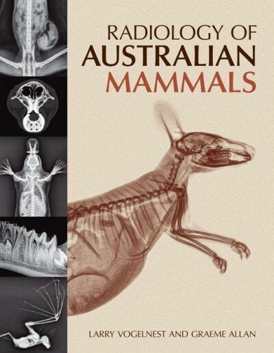
Radiology of Australian Mammals PDF. Interest in the conservation and welfare of Australian native wildlife continues to grow. Veterinarians are frequently presented with injured, diseased or orphaned animals and there is increasing veterinary involvement in conservation programs. In Australia and overseas, Australian mammals are used in research, kept as pets and are popular display and education animals in zoos and fauna parks.
The recognition, diagnosis and treatment of injury and disease in wildlife species present unique challenges for the veterinarian. Radiology is a fundamental diagnostic tool that can be used to further define the nature and extent of injury or disease, guide therapeutic decisions and determine prognosis. An essential aspect of radiology is the recognition and description of abnormal findings. In order to recognise abnormalities, knowledge of normal radioanatomy is necessary. Radiology of Australian Mammals provides a detailed reference on the normal radioanatomy of Australian mammals.
A chapter on radiographic technique covers digital radiography of small species, and restraint and positioning to obtain diagnostic images. This is followed by chapters covering the normal radioanatomy of the short-beaked echidna, platypus, macropods, koala, wombats, dasyurids, possums and gliders, bandicoots and the bilby, and bats. Each chapter includes a detailed description of anatomy relevant to radiography and multiple images of normal radiographs with outlines and annotations identifying relevant structures. A chapter on dental radiology discusses and demonstrates normal dental radioanatomy. The final chapter includes selected radiographic pathology case studies providing an appreciation of radiographic findings seen in some common diseases of Australian mammals. A checklist of the mammals of Australia and its territories and a glossary of abbreviations and terms used for annotation of images complete the volume.
Detailed descriptions of anatomy, highlighting unique features of the taxonomic group
Comprehensively illustrated with detailed, high quality digital radiographs
Illustrates normal radiographic anatomy, allowing veterinary practitioners to identify what is abnormal in their patients
A useful reference for students and zoologists studying anatomy and biology of Australian mammals
Depicts common diseases and injuries that can be visualised radiographically
Table of Contents
Preface
1 Radiographic technique
2 Short-beaked echidna
3 Platypus
4 Macropods (potoroids and macropodids)
5 Koala (co-authored by Dr Susan Hemsley)
6 Wombats
7 Dasyurids
8 Possums and gliders
9 Bandicoots and Bilby
10 Bats
11 Dental radiology (Nadine Fiani)
12 Radiographic pathology case studies (Frances Hulst, Graeme Allan, Larry Vogelnest)
Appendix 1 A checklist of the mammals of Australia and its territories (Paul Andrew)
Index


![Ettinger’s Textbook of Veterinary Internal Medicine 9th Edition [True PDF+Videos]](https://www.vet-ebooks.com/wp-content/uploads/2024/10/ettingers-textbook-of-veterinary-internal-medicine-9th-edition-100x70.jpg "Ettinger’s Textbook of Veterinary Internal Medicine 9th Edition [PDF+Videos]")


