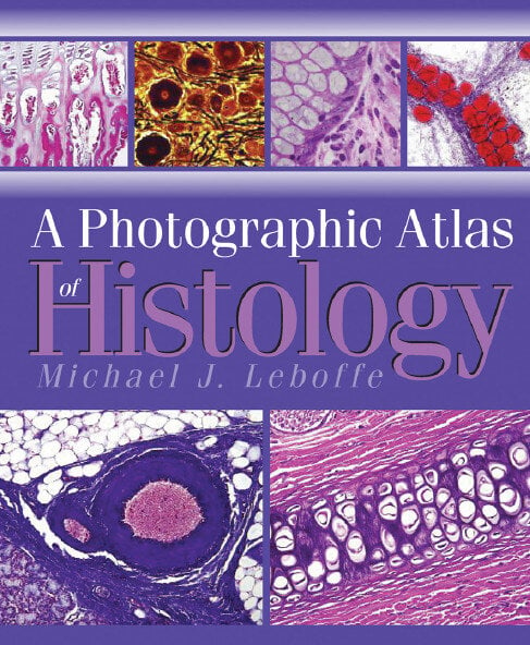
A Photographic Atlas of Histology

By Michael J. Leboffe
A Photographic Atlas of Histology PDF is designed for use in undergraduate histology and human anatomy courses. This atlas contains over 550 high-quality photomicrographs of human tissues and organs.
From Preface:
The primary goal and overriding theme of A Photographic Atlas of Histology is practicality. And while I expect that it will get used during home study and test review, I wrote with the student sitting at the microscope with a box of slides to examine in mind. My hope is that the images in this book will assist that student in identifying what needs to be seen. Toward this end, I used commercially available microscope slides to photograph, so the images represent the quality and diversity of what a student is actually likely to encounter in the laboratory; pathological specimens have not been used. I also have minimized the inclusion of electron micrographs, because beginning histology students are not typically required (or even allowed) to use an electron microscope. Finally, I wrote captions for the images as if I were showing projected images to a class, so they tend to be lengthy and descriptive.
| File Size | 18.7 MB |
| File Format | |
| Download link | Free Download | Become a Premium, Lifetime Deal |
| Support & Updates | Contact Us | Broken Link |
| Join Our Telegram Channel |  |
| More Books: | Browse All Categories |


![Ettinger’s Textbook of Veterinary Internal Medicine 9th Edition [PDF+Videos] Ettinger’s Textbook of Veterinary Internal Medicine 9th Edition [True PDF+Videos]](https://www.vet-ebooks.com/wp-content/uploads/2024/10/ettingers-textbook-of-veterinary-internal-medicine-9th-edition-100x70.jpg)

![Textbook of Veterinary Diagnostic Radiology 8th Edition [PDF+Videos+Quizzes] Thrall’s Textbook of Veterinary Diagnostic Radiology, 8th edition PDF](https://www.vet-ebooks.com/wp-content/uploads/2019/09/textbook-of-veterinary-diagnostic-radiology-8th-edition-100x70.jpg)


