
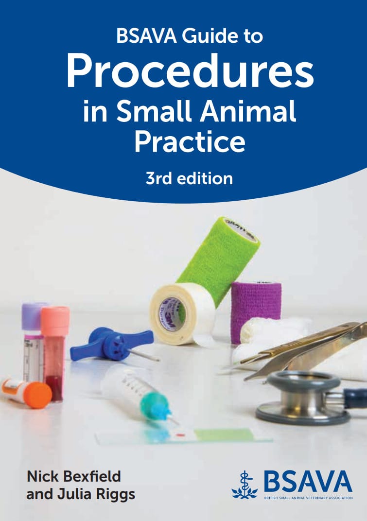
By Nick Bexfield and Julia Riggs
BSAVA Guide to Procedures in Small Animal Practice, 3rd Edition combines high-quality imagery and illustrations, access to a suite of procedural videos and clear, comprehensive step-by-step techniques to provide an essential resource for diagnostic and therapeutic procedures in small animal practice. All procedures have been fully reviewed and updated, and four new procedures have been added, including cystostomy tube placement, endotracheal intubation, point-of-care ultrasound (POCUS) and wet-to-dry dressings. With alphabetised procedures, clear layout and references to further information, the third edition of the Guide is a valuable reference for vets, vet nurses, graduates and experienced clinicians alike.
This Book is Avaiable for Members
Access this book instantly with a Premium membership
4 Essential Vet Clinic Layout Considerations To Improve Your Client’s Experience


Targeting clients’ satisfaction and creating a wide client base requires aligning with the service level they expect to get in your clinic. Your veterinary clinic layout must consider their perceptions about the convenience and comfort you provide your clients with, as well as their needs and first impressions.
It’s not only restricted to the clinical result of their visits, your expertise and accuracy as a veterinarian can do nothing without a comfortable and stress-free environment for both clients and their pet animals enhancing the potentiality for further visits and consults.

Planning for a veterinary clinic layout
Logically, the pet animal will never come without its owner, and each owner expects a specific level of service, comfort, privacy and safety they will find in your pet clinic.
Considering clients’ impressions of the comfort they find in the exterior in addition to the first impression of their visit, you should consider the exterior aspect of your veterinary clinic layout.
Learn how your client care impacts your veterinary practices.
Considerations for Veterinary Clinic Layout
Clients’ experience in your clinic depend on external and internal details that immediately affect their impressions starting from arrival to the building, entrance, going through the reception and ending with sitting in the waiting area.
Here are recommendations for each station:
Arrival
It’s a point of increasing thoughts and perceptions, and here are some considerations and tips that reflect the quality of the service and comfort you provide your clients with:
Location
-You shall consider how easy your client can find the clinic and how obvious the entrance is.
Car Parking
-A location with an available area for cars to park would be a good choice.
-Using parking signs and lines to help find spaces as possible and signs as guidance to no parking or secured areas.
-Places for wheelchairs to be loaded from cars will be useful.
Light
-Showing the entrance at night times, guiding at the entrance passage and car parking reflects how you care for your clients.
Wheelchair Ramp
-Ensuring easy access for owners with mobility challenges and a hint of respecting and welcoming all pet owners.
Green Area
-Green walking spaces for owners and their pet animals to walk and relieve stress before admission.
-Designing the garden with litter boxes and bins can be useful to minimize the probability of urinating or defecating in the waiting area.
Entrance
Presuming a variety of scenario cases which require initiative and immediate assistance can help you with a better veterinary clinic layout.
Your client may be a mother carrying her baby, maybe an elder with a wheelchair, or may come with two different animals or even two or more different species.
Your clients aren’t on the same level of controlling their animals and a conflict between two dogs or escaping shall be also assumed.
Criteria of Ideal Entrance:
- Transparent glass will be the best choice to reflect what’s going on the opposite side.
- A large door with two parallel approaches is preferred; the left one as an ingress and the right one as an egress to minimize the probability of congestion and overstocking at the entrance.
- A sufficient space between the door and the reception counter shall be also considered to avoid interrupting the ingress passage.
- A manually opened door is preferred.
Reception
Reception’s layout affects the performance of reception employees, and clients’ first impression depends on how smoothly and quickly their enquiries and needs are received and answered.
Greeting clients, eye contact and interacting with owners in the waiting area shall be also considered in the veterinary clinic layout.
Keeping the safety of your employees in mind, receptionists are more exposed to the risk of aggressive clients or robbery attempts.
Here are tips to ensure a convenient reception area for both receptionist and clients:
Position
A desk with a suitable seat that gives easy access to a computer, keyboard, printer and credit card machine ensures smooth and quick performance.
Location
Preferred an obvious and easily reached for clients in both the inside and outside areas.
Access to waiting areas
which enables direct interaction with owners and maintains items displayed for sale.
Counter’s Height
Your clients are not always the same, a child or a client with a wheelchair finds difficulty interacting with the receptionist, so, it shall accommodate different points to serve clients and show respect in various conditions.
Access To The Clinical Area
Access to the clinical team reinforces communications which in one way or another affects the whole performance. But unlimited access to dispensary areas isn’t preferred.
Security
A desk linking back to an area of colleagues enhances receptionists’ safety.
Waiting Area
Well well-planned veterinary clinic layout gives priority to clients’ experience during the short period before the appointment.
A comfortable, quiet and stress-free waiting area improves your image.
Here are some recommendations to maximize the convenience of waiting:
-Different separated seats to enhance the feeling of comfort and privacy
-One-species areas can be applied to reduce the incidence of conflicts
-Allocating a separate area for noisy dogs, especially for owners seems to be unsocial
Learn more about Making your veterinary practice a client-friendly space.
Conclusion
Targeting your clients helps you know more about their needs, thoughts and perceptions that you in no way can neglect in your veterinary clinic layout.
Your client’s satisfaction isn’t restricted only to your expertise or accuracy as a veterinarian, they also follow their perception to get a certain level of convenience and comfort to get in your clinic starting from arrival and how obviously the clinic is located passing through the reception and how they are handled and the how comfort they find the waiting area.
Each station has considerations to be kept in mind to reinforce your client’s experience.
Do You Want To Increase Your Veterinary Knowledge and Practical Skills?
You Can Now Browse and Download +3000 Veterinary Books Online In All Veterinary Fields.
Clinical Anatomy and Physiology for Veterinary Technicians 4th Edition

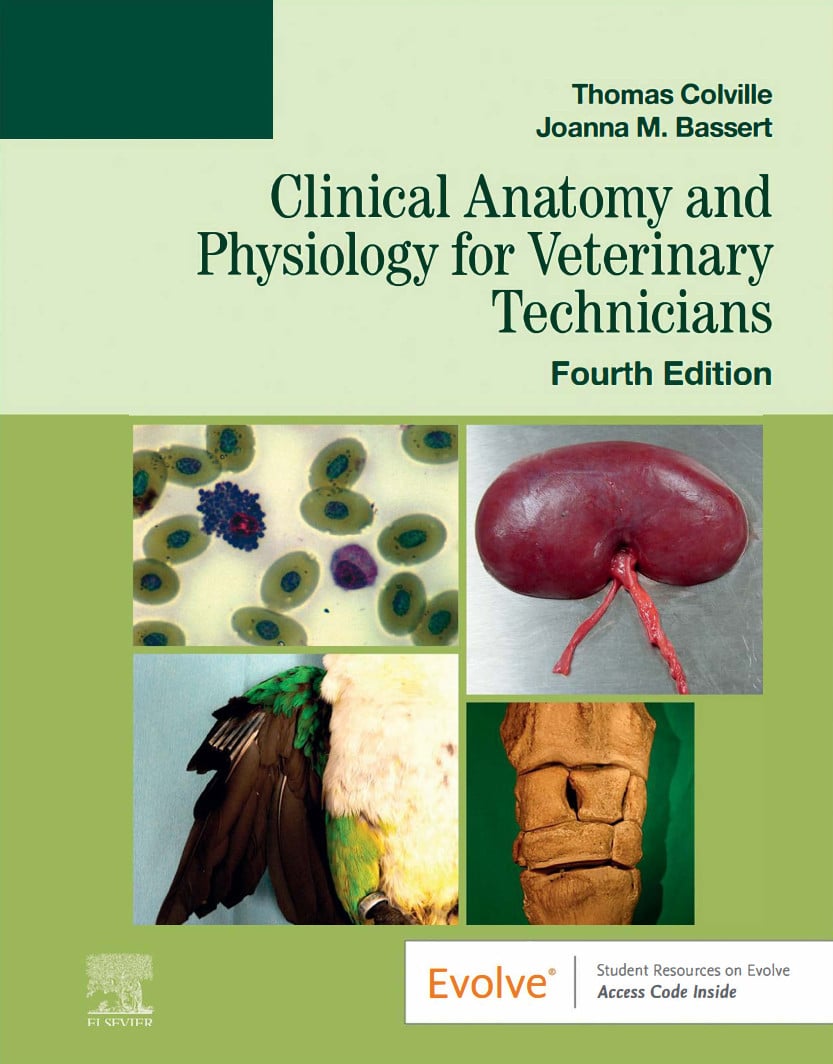
By Thomas P. Colville and Joanna M. Bassert
Clinical Anatomy and Physiology for Veterinary Technicians 4th edition provides the fundamental anatomical and physiological principles necessary for professional success. This textbook not only provides an extensive examination of the various cellular and systemic functions of animal bodies, but also incorporates numerous practical exercises, vocabulary lists, and self-assessment questions throughout each chapter to aid students in developing a solid understanding of anatomic structure and its practical implications.
This Book is Avaiable for Members
Access this book instantly with a Premium membership
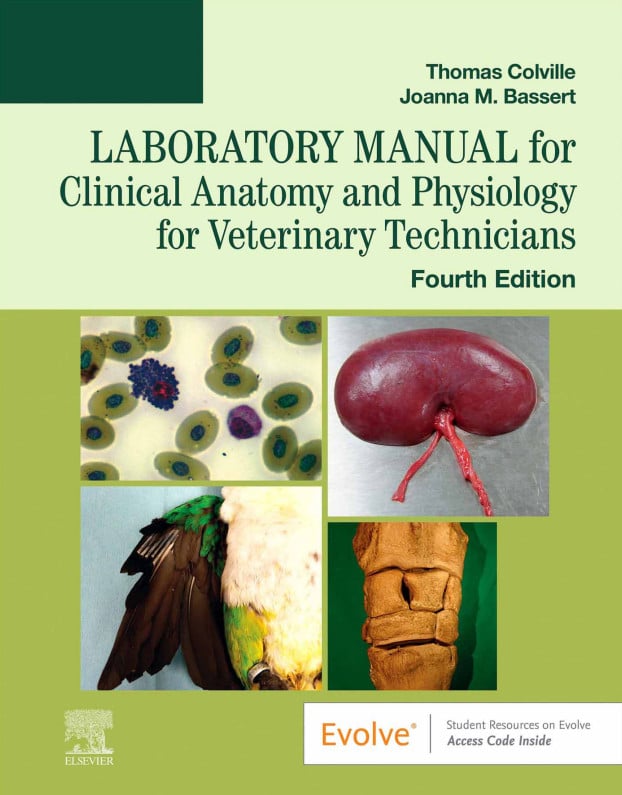
Laboratory Manual for Clinical Anatomy and Physiology for Veterinary Technicians, 4th Edition
By Thomas Colville and Joanna Bassert
Laboratory Manual for Clinical Anatomy and Physiology for Veterinary Technicians, 4th Edition features a variety of activities, such as terminology exercises, illustration identification and labelling, case presentations, and more to help reinforce your understanding of veterinary anatomy and physiology. The laboratory manual also features vivid illustrations, lists of terms and structures to be identified, and step-by-step dissection guides to walk you through the dissection process.
This Book is Avaiable for Members
Access this book instantly with a Premium membership
A Professional’s Guide to Feline Behaviour: Understanding, Improving and Resolving Problems

By Caroline Clark
A Professional’s Guide to Feline Behaviour: Understanding, Improving and Resolving Problems is an invaluable resource for the feline professional, yet written in such an accessible way that it would be of interest to anyone who shares their life with a cat and wants to gain a deeper understanding of their behaviour.
A Professional’s Guide to Feline Behaviour is presented in easy-to-navigate sections, each packed with practical advice and the colour illustrations, tables and graphics throughout make it approachable for every type of reader.
Written by Caroline Clark, a Registered Clinical Animal Behaviourist and RCVS listed veterinary nurse, this well-researched book draws from her knowledge and professional experiences, offering a unique insight into feline behaviour.

This Book is Avaiable for Members
Access this book instantly with a Premium membership
A Guide to Managing Zoo Animal Welfare: A Behavioral Approach

By Bethany L. Krebs and Jason V. Watters
A Guide to Managing Zoo Animal Welfare: A Behavioral Approach delivers a step-by-step guide to behavioral assessment approaches, techniques, and tools for animal welfare with an emphasis on animals living in zoos and aquaria. The authors develop a unique “balance-based” approach that can be used to assess and enhance the welfare of a diverse range of species. Backed by extensive scientific literature, this book also provides foundational context to help readers to understand why the authors give these recommendations and guidelines.

This Book is Avaiable for Members
Access this book instantly with a Premium membership
Laboratory Manual for Laboratory Procedures for Veterinary Technicians, 8th Edition

By Kristin J. Holtgrew-Bohling
Reinforce the essential information you need to master with the Laboratory Manual for Laboratory Procedures for Veterinary Technicians, 8th Edition. Corresponding to each unit in Laboratory Procedures for Veterinary Technicians, 8th Edition, this full-color manual includes various exercises and test questions that help you focus on learning key concepts and skills for the veterinary clinical setting. Fill-in-the-blank exercises, lab exercises, crossword puzzles, word searches, photo quizzes, lab forms, specimen pictures, and review questions all help to clarify more challenging concepts. Plus, this edition includes content on fear-free handling, specimen collection methods, immunology, and quality control.

This Book is Avaiable for Members
Access this book instantly with a Premium membership
Assessing Essential Skills of Veterinary Technology Students 4th Edition

By Lisa E. Schenkel, Amanda Colón, Sandra Lynn Bertholf, Sabrina Timperman and Laurie J. Buell
Assessing Essential Skills of Veterinary Technology Students 4th Edition provides a comprehensive review of the required American Veterinary Medical Association Committee on Veterinary Technician Education and Activities (AVMA CVTEA®) essential skills for completion of a veterinary technology degree. Each essential skill includes assessment criteria as well as decision-making instructions necessary to demonstrate proficiency both academically and professionally.
Assessing Essential Skills of Veterinary Technology Students is organized based on the categories provided by the AVMA CVTEA, making it easy for an instructor and students to locate the assessment criteria for a particular essential skill relative to their course.

This Book is Avaiable for Members
Access this book instantly with a Premium membership
Antimicrobial Therapy in Veterinary Medicine 6th Edition

By Patricia M. Dowling, John F. Prescott and Keith E. Baptiste
Antimicrobial Therapy in Veterinary Medicine 6th Edition has been updated to reflect advances in the field, including new international contributors and a broader global outlook. It includes extensive knowledge of both general principles of mechanisms of antimicrobial drug action including specific classes of antimicrobial agents, as well as chapters dedicated to antimicrobial drug use in a wide range of animal species. As antimicrobial resistance increases as a major global issue in both human and animal health, this book’s renewed focus on antimicrobial stewardship in companion animals, in food animals, and on global aspects keeps it at the forefront of this vital field.

This Book is Avaiable for Members
Access this book instantly with a Premium membership
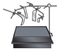
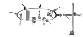
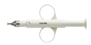
Veterinary Surgical Instruments List: Names and Pictures

As a Completion To the Previous Article about Veterinary Equipment and Tools List, In this blog post, we will take a look at some common Veterinary Surgical Equipment List With Names, Uses, and Pictures in veterinary clinics.
Veterinary surgery is an important branch of veterinary medicine, we can’t begin learning veterinary surgery without knowing all the surgical tools you will use in any surgery that’s why we make this Veterinary Surgical Instruments List for all veterinary students to know all Veterinary Surgical Instruments List or veterinarians have come across the issue of needing to purchase surgical instruments for clinics.
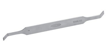
General Surgical Instruments
General veterinary surgical instruments are the basic tools used in all operations. These instruments must be manufactured with high precision and years of experience to ensure the best results. Similarly, the user must be well-trained and experienced to achieve optimal surgical outcomes.
Below are commonly used general instruments in veterinary surgery:
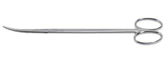
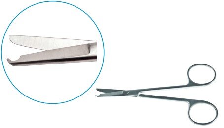
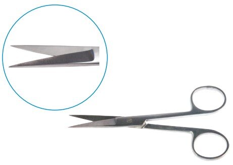
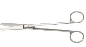
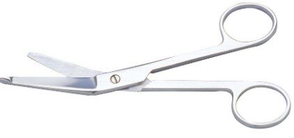
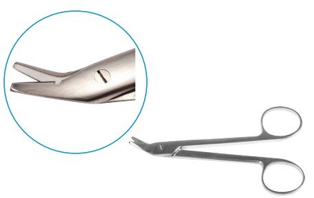
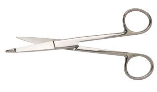
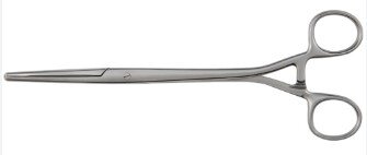
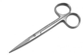
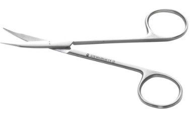
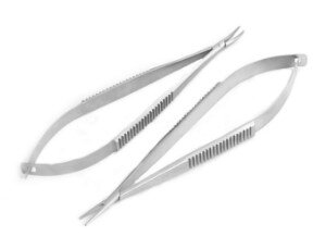
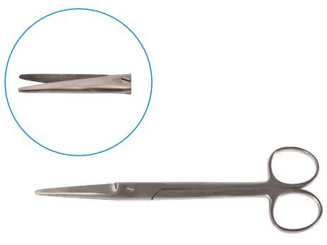
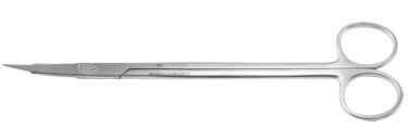
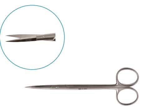
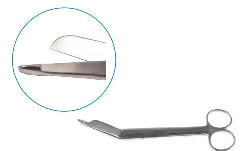
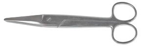
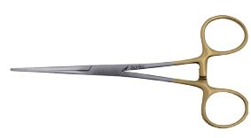
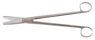
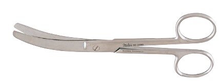
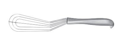
Scissors
Scissors are essential tools used by surgeons to cut and dissect tissues, muscles, organs, and sutures. They allow for the fast and safe removal of obstructive tissues during procedures. Made from balanced stainless steel, surgical scissors are thinner, sharper, and pointed for precision cutting.
| Instrument | Use | Image |
|---|---|---|
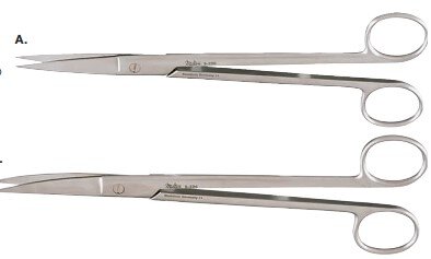
| Mayo | Soft tissue cutting | 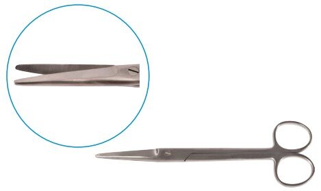 |
| Metzenbaum | Soft tissue cutting |
 |
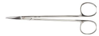
| Spencer Stitch | Suture removal |
 |
| Standard | Suture removal |
 |
| Careless | Suture removal |
 |
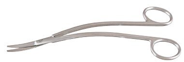
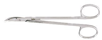
| Lister | Bandage cutting |
 |
| Wire Scissors | Cut stainless steel wire |
 |
| Knowles Bandage Scissors | Remove bandage and dressing |
 |
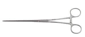
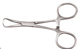
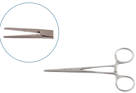
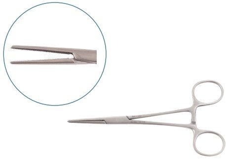
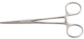
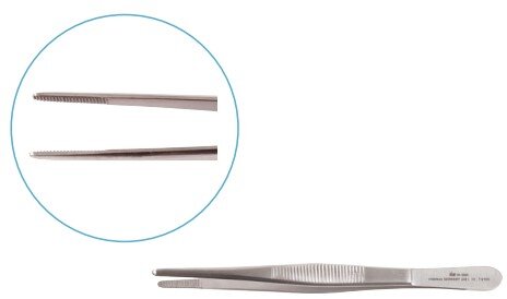
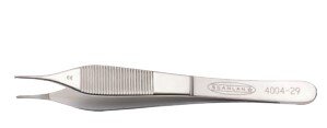
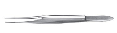
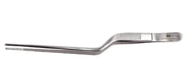
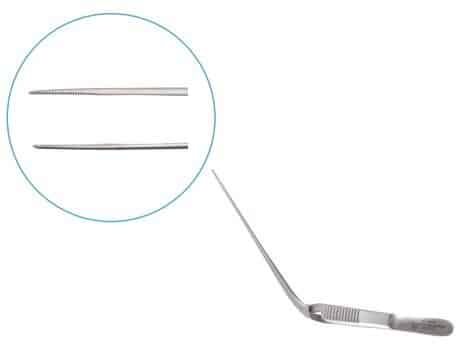
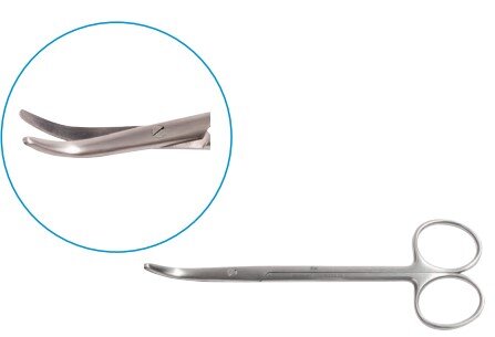
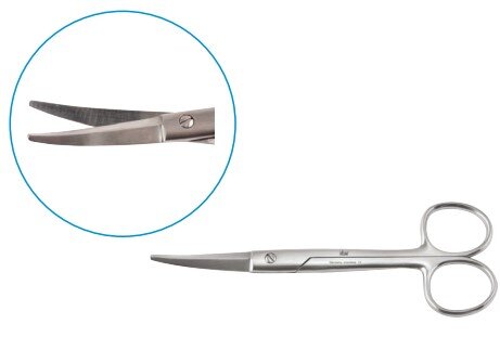
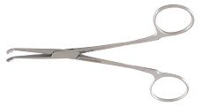
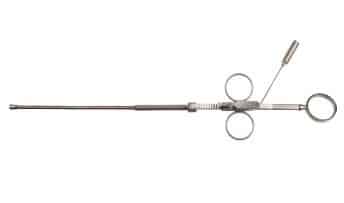
Forceps
Forceps are essential surgical tools used to hold tissues, separate structures, improve access, and assist in cutting and suturing. They are also used for tweezing and applying pressure during procedures.
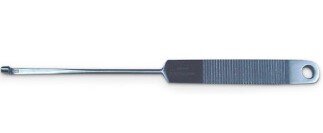
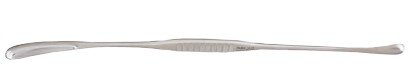
Dissecting Forceps
Dissecting forceps are temporary tools used to handle skin, tissues, and organs during surgery. Surgeons also use them to guide needles or manipulate delicate structures.
| Type | Use | Image |
|---|---|---|
| Standard plain & toothed | Handle soft tissues | 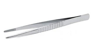 |
| Mosquito | Hold small capillaries | 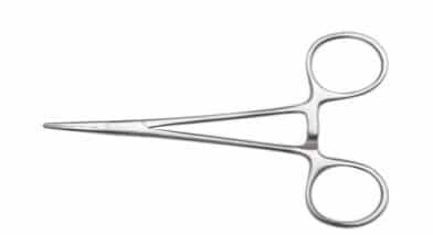 |
| Adsons | Handle soft tissues | 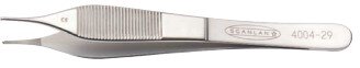 |
| Continental Standard | Handle skin | 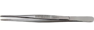 |
| Emmett | Handle deep tissues | 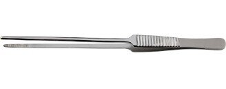 |
| Debakey | Handling of viscera | 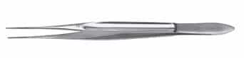 |
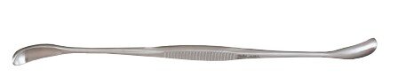
Tissue Forceps
Tissue forceps are used for delicate manipulation of tissues without causing trauma. They provide secure grip and precise handling of soft organs and structures during surgical procedures.
| Type | Use | Image |
|---|---|---|
| Babcock | Handling viscera and soft tissue | 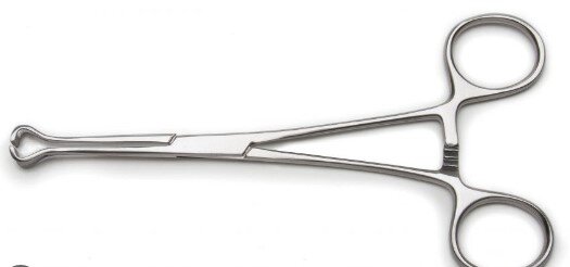 |
| Allis | Handling soft tissue | 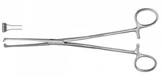 |
| Duval | Handling viscera and tissue | 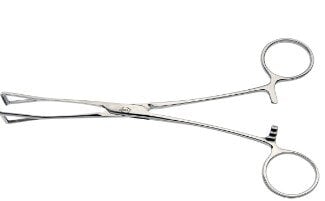 |
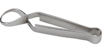
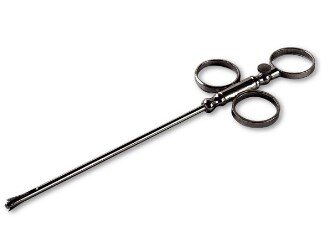
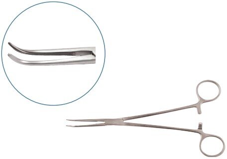
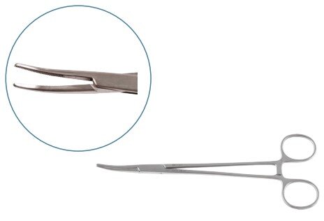
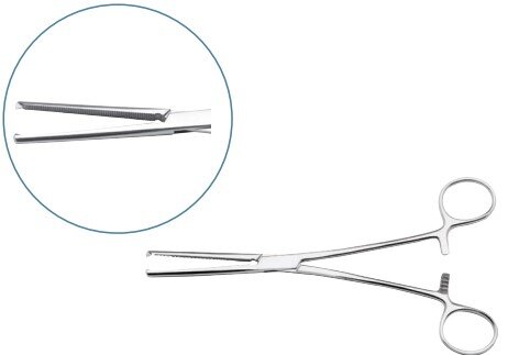
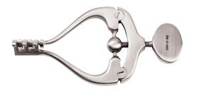
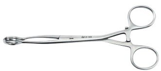
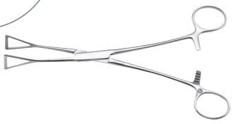
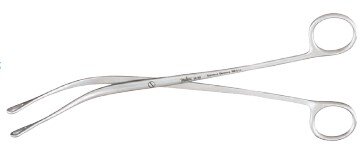
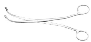
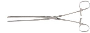
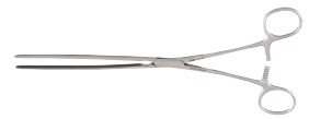
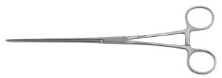
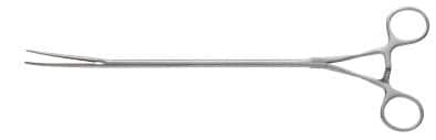
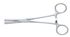
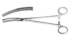
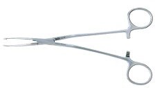
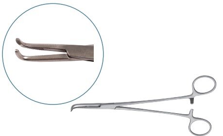
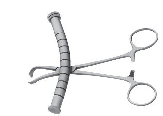
Clamps or Hemostats
Hemostats are essential surgical instruments used to occlude blood vessels either completely or partially. They help control bleeding during surgery and are commonly used to block blood flow to internal organs temporarily to protect them during procedures.
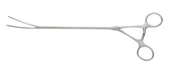
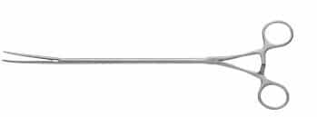
Visceral Clamps
Used to occlude visceral organs like the stomach, intestines, or cervix.
| Type | Use | Image |
|---|---|---|
| Doyen Mayo Robinson | Occlusion of stomach and intestine |
 |
| Parker Kerr | Occlusion of cervix |
 |
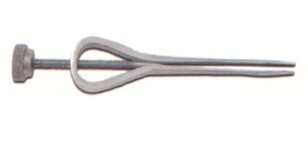
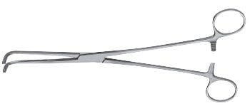
Towel Clamps
Used for securely attaching surgical drapes to the patient’s skin.
| Type | Use | Image |
|---|---|---|
| Cross Action | Attach drapes to the surgical area |
 |
| Backhaus | Attach drapes to the surgical area |
 |
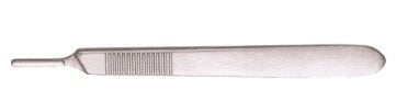
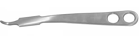
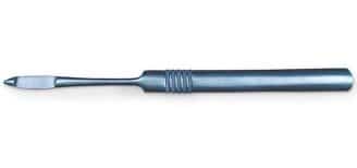
Scalpel
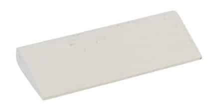
The scalpel is a long, thin surgical instrument used to make precise cuts in the skin and underlying tissues. It is an essential tool in dermatology and surgery, enabling incisions, tissue dissections, and various surgical techniques. The term “scalpel” originates from Latin, meaning “small knife.” Scalpels come in a range of sizes and shapes, each suited for specific procedures.
| Type | Use | Image |
|---|---|---|
| Scalpel Handle | Hold surgical blades |
 |
| Scalpel Blades | Make incision and tissue transection |
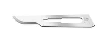 |
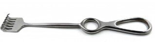
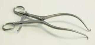
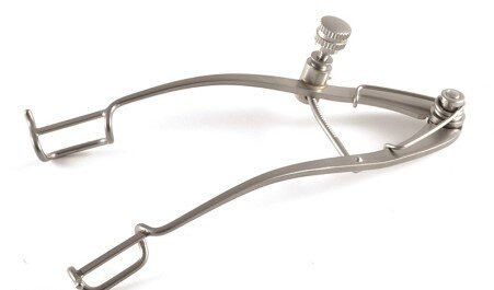
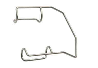
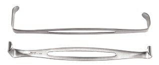
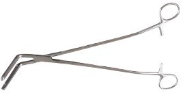
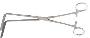
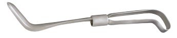
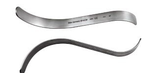
Retractors
Retractors are used to hold open the incision or wound during surgery, allowing the surgeon better visibility and access to the surgical site. They help retract soft tissues, joints, or organs out of the way, and can be manually held or self-retaining to free the surgeon’s hands.
Manual Retractors
These retractors require manual handling to maintain exposure of tissues and surgical fields.
| Type | Use | Image |
|---|---|---|
| Langenbeck | Soft tissue retraction |
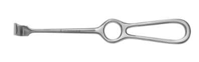 |
| Volkman | Retraction of tendons and muscles |
 |
| Hohman | Retraction of joints |
 |
| Czerny | Soft tissue retraction |
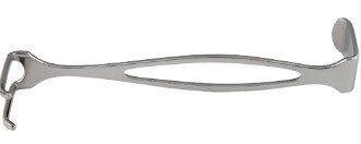 |
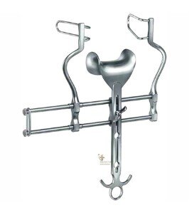
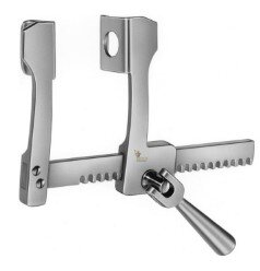
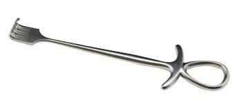
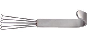
Self-Retaining Retractors
These retractors do not require constant holding and are designed to hold the tissue apart automatically, keeping the surgical field open.
| Type | Use | Image |
|---|---|---|
| Gelpi | Joint and muscle retraction |
 |
| Cone | Used in orthopedic surgeries |
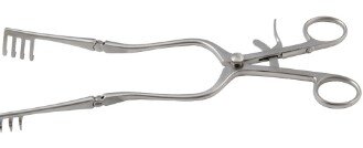 |
| Travers | Joint and muscle retraction |
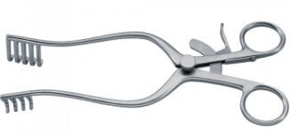 |
| Gosset | Abdominal wall retraction |
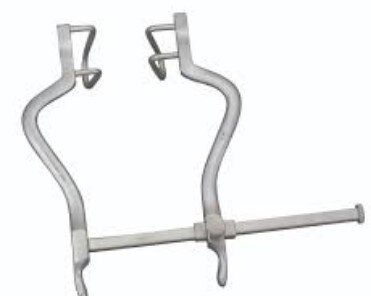 |
| Balfour | Liver retraction |
 |
| Finochietto | Spread ribs |
 |
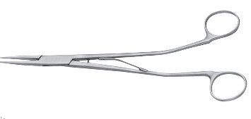
Needle Holder
A needle holder, also known as a needle driver, is a surgical instrument designed to securely hold suturing needles. It features a comfortable handle for one-handed use and a shaft with a tip that guides the needle through tissues with precision. Some types also incorporate built-in scissors for cutting sutures.
| Type | Use | Image |
|---|---|---|
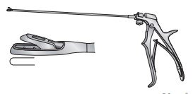
| Gillies | Hold needle and cut suture | 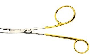 |
| Olsen Hegar | Hold needle and cut suture | 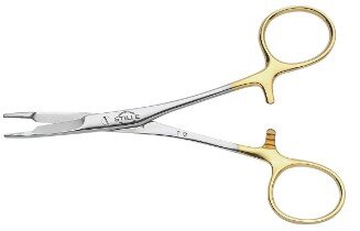 |
| Mayo Hegar | Hold needle | 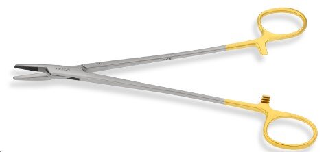 |
| Bruce Clarke | Hold needle | 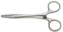 |
| McPhail | Hold needle | 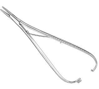 |
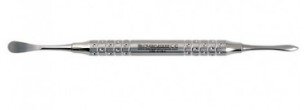
Specific Surgical Instruments
The surgical instruments used by veterinarians during operations must be of the highest quality due to the fragility of organs and bones. Even minor negligence can lead to severe damage. That’s why top-grade, specially designed tools are crucial in veterinary practice.
Orthopedic Surgical Equipment
Orthopedic instruments are used by surgeons to diagnose and treat bone fractures, cut or remove bone, and perform orthopedic procedures. These tools are precision-crafted for efficient and safe handling of skeletal structures.
| Instrument | Use | Image |
|---|---|---|
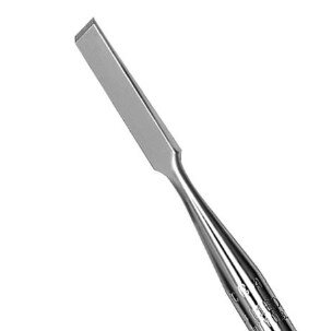
| Chisel | Bone shaving |
 |
| Gouge | Bone shaving |
 |
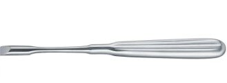
| Osteotome | Precise bone cut |
 |
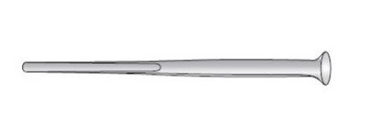
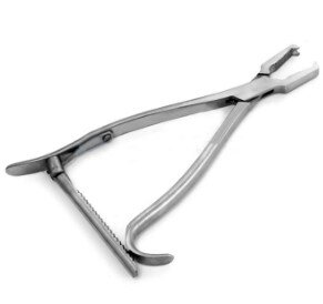
| Periosteal Elevator | Raise periosteum before drilling |
 |
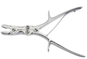
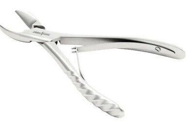
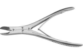
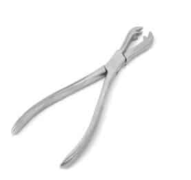
| Rongeurs | Nibble bone pieces |
 |
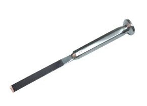
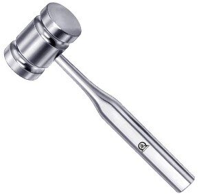
| Mallet | To use with chisel |
 |
| Paton Bone Cutting Forceps | Cut bone |
 |
| Ruskin Liston Bone Cutting Forceps | Cut bone |
 |
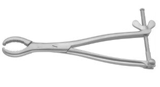
| Ferguson Bone Holding Forceps | Prevent bone movement in surgery |
 |
| Kern Bone Holding Forceps | Prevent bone movement in surgery |
 |
| Hey Groove Bone Holding Forceps | Maintain tip closure |
 |
| Jacob Chuck | Insert and remove pins (intramedullary pinning) |
 |
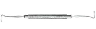
Ophthalmic Surgical Instruments
Ophthalmic surgical tools are used for carrying out eye-related surgeries. Both cornea and lens-related surgeries are done with the help of different surgical instruments.
Scissors
| Instrument | Use | Image |
|---|---|---|
| Iris scissors | To cut iris |
 |
| Castroviejo scissors | Cut lens capsule |
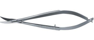 |
| Tenotomy scissors | For fine dissection |
 |
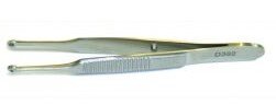
Forceps
| Instrument | Use | Image |
|---|---|---|
| Chalazion forceps | Stabilize eyelid and protect globe |
 |
| Benet cilia forceps | For plucking eyelashes |
 |
| Catford forceps | Help in risk-free suturing |
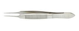 |
| Capsulorhexis forceps | Grasp lens capsule |
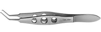 |
| Micro corneal tying forceps | Tie suture material |
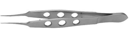 |
| Capsule forceps | Grabs lens capsule |
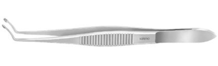 |
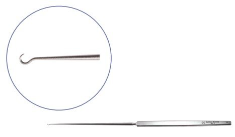
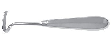
Hooks
| Instrument | Use | Image |
|---|---|---|
| Kirby expressed hook | Use for lens removal |
 |
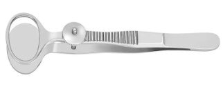
Speculums
| Instrument | Use | Image |
|---|---|---|
| Williams speculum | Provide access to eyeball by a retraction |
 |
| Barraquer speculum | Provide access to eyeball by a retraction |
 |
Dilators
| Instrument | Use | Image |
|---|---|---|
| Nettleship dilator | Dilate narrow canals |
 |
Needle Holder
| Instrument | Use | Image |
|---|---|---|
| Castroviejo needle holder | Hold needle during suturing |
 |
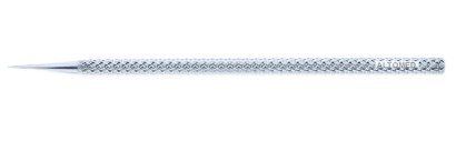
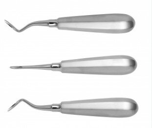
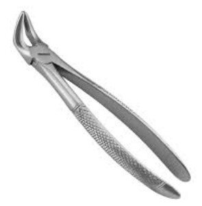
Dental Instruments
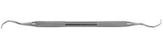
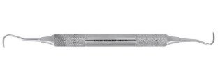
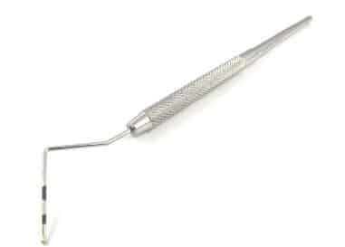
Dental instruments are very important for every single dental surgery. They are the most common tools used by vets for the diagnosis and treatment of oral problems—such as removing dental calculus, repairing teeth, and performing extractions. Instruments like scalers, elevators, curettes, and chisels each serve specific functions. Below are commonly used dental surgical instruments:
| Instrument | Use | Image |
|---|---|---|
| Dental elevator | To separate connection of teeth and bone |
 |
| Extraction forceps | Extract tooth |
 |
| Periosteal elevator | To expose bone |
 |
| Subgingival curette | Remove the unwanted material from the mouth |
 |
| Dental explorer | Expose the hard surface of teeth |
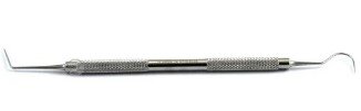 |
| Supragingival scalers | Remove supragingival calculus |
 |
| Periodontal probe | Measure depth of periodontal pocket |
 |
| Sharpening stone | For sharpening instruments |
 |
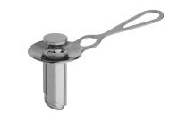
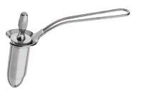
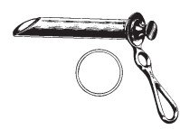
Teat Instruments
Cows play a crucial role in dairy farming by providing milk, an essential dietary component. However, their teats are prone to damage or infections, which can hinder milk production. Teat surgical instruments are used to treat these issues effectively and maintain udder health. Below are common teat surgical tools:
| Instrument | Use | Image |
|---|---|---|
| Dilator | Open teat canal |
 |
| Slitter | Clear the teat canal by incision from inside to outside |
 |
| Tumor extractor | Remove fibrous material from teat canal |
 |
| Udder infusion canula | Administer medicine in teat canal |
 |
| Teat curette | Clean inside of teat canal |
 |
| Lichty teat knife | Open stenotic teats |
 |
| Milking tubes | Keep injured teat open |
 |
Plastic Surgery Instruments
Plastic surgery is a form of body modification that can be approached for a myriad of reasons – whether it is for someone who wants to improve their appearance or for someone who needs to reconstruct parts of their body that have been damaged by injury or disease…
Clamps
| Instrument | Function | Image |
|---|---|---|
| Kelly clamp | Hold heavy tissues |
 |
| Crile clamp | Hold suture ends |
 |
| Rankin clamp | Clamp tissues for ligation |
 |
| Rochester Pean clamp | Clamp larger vessels |
 |
| Moynihan clamp | Suturing of vascular tissues |
 |
| Schmidt clamp | Suturing stalk of tissues |
 |
| Kocher clamp | Grasp heavy tissues |
 |
Forceps
| Instrument | Function | Image |
|---|---|---|
| Dressing forceps | Dressing wounds |
 |
| Adson forceps | Hold dressing material |
 |
| DeBakey forceps | Avoid tissue damage |
 |
| Jansen forceps | Remove boney septum |
 |
| Wilde forceps | Stop nasal bleeding |
 |
Scissors
| Instrument | Function | Image |
|---|---|---|
| Mayo scissors | Cut tissue and suture |
 |
| Martin scissors | Cut cartilage |
 |
| Joseph scissors | Cut tissues and sutures |
 |
| Forman scissors | Dissect nasal cartilage |
 |
| Aufrect scissors | Cut delicate tissues |
 |
| Lister bandage scissors | Cut dressing and bandages |
 |
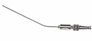
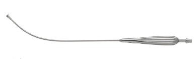
Evacuation Instruments
| Instrument | Function | Image |
|---|---|---|
| Frazier suction tube | Remove debris and fluid from surgical spaces |
 |
| Adson suction tube | Aspire blood and residues |
 |
| Baron suction tube | Remove fluid and debris |
 |
Retractor and Exposure
| Instrument | Function | Image |
|---|---|---|
| Army Navy retractor | Retract bone and skin |
 |
| Murphy retractor | Retract delicate tissues |
 |
| Bear claw retractor | Help in a facelift |
 |
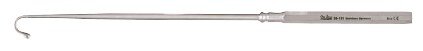
| Gilles skin hook | Retract skin |
 |
| Jansen retractor | Spread tissues |
 |
Gastrointestinal Instruments
The digestive system is an important part of the body! It’s responsible for taking in nutrients from your food and turning it into energy. The upper GI tract (esophagus, liver, stomach, gallbladder, spleen, pancreas, diaphragm) and the lower GI tract (small and large bowel, mesentery, appendix, rectum, omentum, anus) each require specialized surgical instruments. Upper‑GI tools are medium‑length; lower‑GI tools are long or extra‑long.
Cholecystectomy Instruments
| Instrument | Function | Image |
|---|---|---|
| Collin gallbladder forceps | Holding and removal of gallstones |
 |
| Lovelace gallbladder forceps | Grab gallstones |
 |
| Mixter gallstone forceps | Manipulate stone with precision |
 |
| Mayo gallstone scoop | Scoop out gallstone |
 |
| Ferguson gallstone scoop | Remove stones from gallbladder |
 |
| Oschner trocar | Drain fluids from cavities |
 |
| Kidney stone forceps | Remove stones from bile duct |
 |
Liver & Stomach Surgical Instruments
| Instrument | Function | Image |
|---|---|---|
| Wishbone retractor | Abdominal tissue retraction |
 |
| Bookwalter retractor | Increase access to surgical site |
 |
| Benson pylorus dilator | Grip organs & membranes |
 |
| Esophageal dilator | Dilate esophagus |
 |
| Mayo‑Robson forceps | Hold bowel |
 |
| Mayo‑Noble scissors | Dissect tissues & skin |
 |
| Scudder intestinal clamp | Clamp blood vessels |
 |
Lower GIT Instruments
| Instrument | Function | Image |
|---|---|---|
| Doyen clamp | Atraumatic grasping |
 |
| Dennis clamp | Hold tissues |
 |
| Foss intestinal clamp | Clamp bowel |
 |
| Fehland intestinal clamp | Clamp bleeding site |
 |
| Bainbridge forceps | Lock bleeding vessels |
 |
| Dubois scissors | Deep dissection |
 |
| Busch scissors | Cut umbilical cords |
 |
| Terminal end stapler | Close enterotomies |
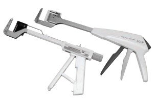 |
| Intraluminal stapler | Seal tissues in colostomy |
 |
| GI anastomosis stapler | Staple transected tube |
 |
Rectal & Anal Instruments
| Instrument | Function | Image |
|---|---|---|
| Fergusen angiotribe clamp | Occlude veins and arteries |
 |
| Buie pile clamp | Grasp hemorrhoids |
 |
| McGivney hemorrhoid ligator | Remove hemorrhoids |
 |
| Miller rectal scissors | Dissect tissues in anal canal |
 |
| Sims scissors | Cut bandages & dead tissues |
 |
| Kelly fistula scissors | Cut tissues |
 |
| Quimby scissors | Cut delicate tissues |
 |
| Yeoman biopsy forceps | Used for diagnosis |
 |
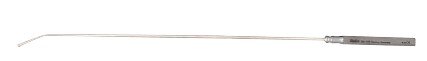
| Buie fistula probe | View anal sac |
 |
| Pratt rectal probe | Remove O‑rings |
 |
| Rosser crypt hook | Remove foreign objects |
 |
| Sawyer retractor | Provide access to lower rectum |
 |
| Hirschman anoscope | Visualize lower rectum |
 |
| Fansler‑Ives anoscope | Examine incision area |
 |
| Hirschman proctoscope | Visual inspection of anal area |
 |
| Rigid sigmoidoscope | Examine inside of anus |
 |
| Chelsea‑Eaton anal speculum | View rectum |
 |
| Pratt rectal speculum | Dilate anal area |
 |
Cardiothoracic And Vascular Surgical Instruments
In the field of cardiothoracic surgery, common procedures include coronary artery bypass grafting and aortic valve replacement. Although approaches and incisions differ, surgical training and expertise are key to outcomes. Advances in medical equipment have given surgeons specialized tools to remove vessels, repair valves, and perform bypasses with greater precision and safety.
Clamps & Related Instruments
| Instrument | Function | Image |
|---|---|---|
| Rochester‑Pean clamp | Clamp larger vessels |
 |
| Kantrowitz clamp | Grasp tissues |
 |
| Finochietto clamp | Grasp tissues & blood vessels |
 |
| Bulldog applicator | Apply bulldog clamp |
 |
| Sweet clip applier | Ligate tubular sutures |
 |
| Gluck rib shears | Cut ribs |
 |
| Doyen rib stripper | Strip periosteum from ribs |
 |
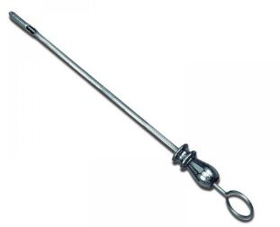
Probes & Dilators
| Instrument | Function | Image |
|---|---|---|
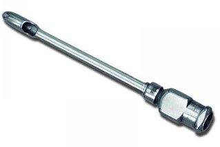
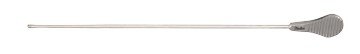
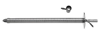
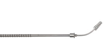
| Garrett vascular dilators | Perform vessel dilation | 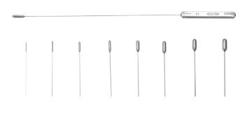 |
Measuring Instruments
| Instrument | Function | Image |
|---|---|---|
| Tessier caliper | Measure anatomical structures | 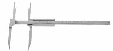 |
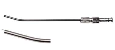
Installation & Evacuation Instruments
| Instrument | Function | Image |
|---|---|---|
| Wolf suction | Remove blood from surgical site |
 |
| Poole suction tube | Remove large volumes of fluid |
 |
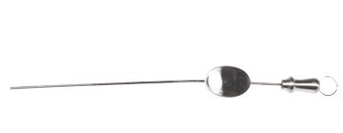
Retraction & Exposure
| Instrument | Function | Image |
|---|---|---|
| Allison retractor | Retract lungs |
 |
| Malleable ribbon retractor | Retract during orbital dissection |
 |
| Leaflet retractor | Used in cardiac surgery |
 |
Special Instruments
| Instrument | Function | Image |
|---|---|---|
| Vessel punch | Create an opening in vessels |
 |
| McIntosh suture holder | Separate sutures |
 |
| Rummel tourniquet | Occlude blood vessels |
 |
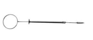
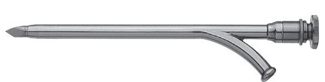
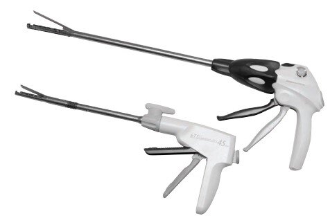
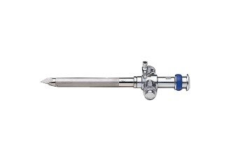
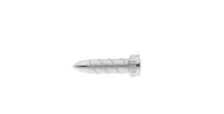
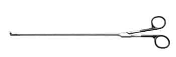
Endoscopic Instruments
Endoscopy is used in animals to examine internal organs via a camera, either diagnostically (viewing the digestive tract through mouth or anus) or therapeutically (removing foreign objects, repairing abnormalities). Under general anesthesia, veterinarians can visualize the heart, lungs, stomach, intestines, and more. Endoscopy improves recovery by avoiding large incisions.
Essential Endoscopic Instruments
| Instrument | Function | Image |
|---|---|---|
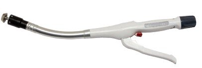
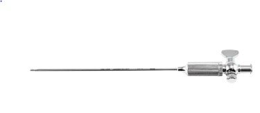
| Endoscopic trocar | Small‑puncture port |
 |
| Veress needle | Establish pneumoperitoneum |
 |
| “S” retractors | Retract abdominal walls |
 |
| Thread sleeve | Reinforce suture connection |
 |
| Balloon dissector | Separate extraperitoneal tissue |
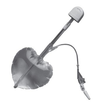 |
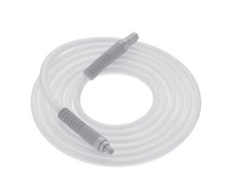
| Insufflation tubing | Insufflate abdominal cavity |
 |
Viewing of Working Space
| Instrument | Function | Image |
|---|---|---|
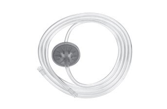
| Fiber‑optic light cable | Illuminate surgical field |
 |
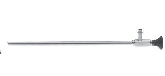
| Telescope | Magnified internal view |
 |
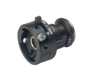
| Camera head | Capture endoscopic images |
 |
| Bipolar cord | Connect bipolar instruments |
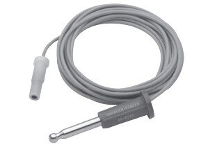
 |
| Monopolar cord | Connect monopolar instruments |
 |
Special Instruments
| Instrument | Function | Image |
|---|---|---|
| Chitwood DeBakey clamp | Clamp lung tissues |
 |
| Dennis clamp | Hold tissues |
 |
| Chitwood suture cutter | Cut sutures |
 |
Other Equipment
Below are additional miscellaneous instruments commonly used in various veterinary surgical procedures.
| Instrument | Use | Image |
|---|---|---|
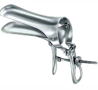
| Cusco Vaginal Speculum | Exposure of vaginal tissue |
 |
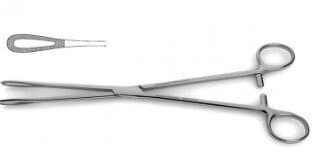
| Rampley Sponge Holding Forceps | Hold swabs and sponges |
 |
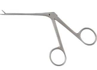
| Hartman Crocodile Forceps | Nasal and oral use |
 |
Conclusion
There are many reasons why surgical instruments are needed in surgeries. First, they are used to cut and pierce the skin. Second, they are used to perform the operation. Third, they are used to remove foreign objects. Fourth, they are used to keep the patient from bleeding. So that’s why we need surgical instruments for surgeries. We hope you enjoyed our article about surgical instruments. You may have never thought about it, but there are a lot of different instruments that are used in surgeries. Understanding the different instruments and what they are used for will help you appreciate the skill and expertise of veterinary surgeons. Since there are so many different instruments used in surgeries, it can be difficult to keep track of them all but we have mentioned the most important ones.
Do You Want To Increase Your Veterinary Knowledge and Practical Skills?
You Can Now Browse and Download +3000 Veterinary Books Online In All Veterinary Fields.


![Ettinger’s Textbook of Veterinary Internal Medicine 9th Edition [True PDF+Videos]](https://www.vet-ebooks.com/wp-content/uploads/2024/10/ettingers-textbook-of-veterinary-internal-medicine-9th-edition-100x70.jpg "Ettinger’s Textbook of Veterinary Internal Medicine 9th Edition")


