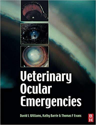
By David L. Williams , Kathy Barrie, Thomas Ffrangcon Evans
Handbook of Veterinary Ocular Emergencies PDF. This practical guide to small animal ophthalmic emergencies is ideal for the non-specialist veterinarian. From a problem-oriented approach, it describes ocular emergencies and gives information for immediate palliative measures and long-term treatment. Clinical pathways, diagnostic flowcharts, bullet points, and easy-to-follow line diagrams provide instant access to the correct diagnosis and management of ocular emergencies. Tinted boxes highlight important issues, key information, and additional material on background pathogenesis and treatment rationale. The emphasis on differential diagnosis and treatment options, as well as recommendations on when to refer a case to a specialist, makes this book an essential consulting room reference for every veterinary practitioner.
Written at an appropriate level for the non-specialist veterinarian, making it a practical guide for managing small animal ophthalmic emergencies.
Provides instant access to the correct diagnosis and management of ocular emergencies with clear, easy-to-use diagnostic flowcharts.
Highlights key information and important issues in tinted boxes throughout the text, making clinical facts accessible to busy practitioners.
Table of Contents
FOREWORD
INTRODUCTORY CHAPTERS
CHAPTER 1: INTRODUCTION
1.1 How to use this book
1.2 Performing an ocular examination in an emergency situation
1.3 Recording observations made in an ocular emergency
1.4 Equipment and aids required to deal with the ocular emergency
1.5 Some preliminary notes on treatment of ocular infections
1.6 Analgesia in ocular emergencies
1.7 Dealing with ocular emergencies in horses and ruminants
1.7.1 Techniques facilitating large animal ocular examination
1.7.2 Techniques facilitating large animal ocular therapeutics
CHAPTER 2: A problem orientated approach
2.1: The red eye
2.2 The painful eye
2.3 The white eye
2.4 The suddenly blind eye
2.5 Ocular lesions in systemic disease
DIAGNOSIS AND TREATMENT OF OCULAR EMERGENCIES
CHAPTER 3: ADNEXA AND ORBIT
3.1: Lid laceration
3.2 Conjunctivitis
3.3 Conjunctival foreign body
3.4 Acute keratoconjunctivitis sicca
3.5 Orbital cellulitis
3.6 Orbital space occupying lesion
CHAPTER 4: GLOBE
4.1: Blunt trauma to the globe
4.2: Globe prolapse
4.3: Penetrating globe injury
CHAPTER 5: CORNEA
5.1: Corneal ulceration
5.1.1: Is an ulcer present? – the use of ophthalmic stains
5.1.2: Three key questions regarding any corneal ulcer
5.1.2.1 Ulcer depth
5.1.2.2 Ulcer healing
5.1.2.3 The cause of the ulcer
5.2 Dealing with different ulcers
5.2.1 The simple healing superficial ulcer
5.2.2 The recurrent or persistent non-healing superficial ulcer
5.2.3 Ulceration secondary to bullous keratopathy
5.2.4 Partial thickness stromal ulceration
5.2.5 Near-penetrating ulcers, descemetocoeles and penetrating ulcers
5.2.6.1 The melting ulcer: diagnosis
5.2.6.2 The melting ulcer: diagnosis
5.3 Corneoscleral laceration
5.3.1 Defining the extent of a corneal laceration
5.3.2 Defining involvement of other ocular structures
5.3.3 Repairing a simple non-penetrating corneal laceration
5.3.4 Repairing a simple perforating corneal laceration
5.3.5 Repairing a corneal laceration complicated by iris inclusion
5.4 Corneal foreign bodies
5.4.1 Recognising a corneal foreign body
5.4.2 Dealing with a non-perforating corneal foreign body
5.4.3 Dealing with a fully penetrating corneal foreign body
5.5 Antibiotics and mydriatic cycloplegia in corneal emergencies
CHAPTER 6: IRIS
6.1 Iritis
6.1.1 Diagnosis: clinical signs
6.1.2 Diagnosis: diagnostic tests
6.1.3 Treatment: pain relief
6.1.4 Treatment: anti-inflammatory medication
6.1.5 Treatment: reducing miosis and preventing synechia formation


![Ettinger’s Textbook of Veterinary Internal Medicine 9th Edition [True PDF+Videos]](https://www.vet-ebooks.com/wp-content/uploads/2024/10/ettingers-textbook-of-veterinary-internal-medicine-9th-edition-100x70.jpg "Ettinger’s Textbook of Veterinary Internal Medicine 9th Edition [PDF+Videos]")


