back to top

โŒ› Limited-Time Offer: Get Lifetime Membership For ONLY $199.99 $149.99 - Deal Will End Soon:

00Hrs 00Mins 00Secs
Sign Up Now
HomeAnimals BooksSmall AnimalsCanine and Feline Cytopathology: A Color Atlas and Interpretation Guide 4th Edition

Canine and Feline Cytopathology: A Color Atlas and Interpretation Guide 4th Edition

-

Canine And Feline Cytopathology: A Color Atlas And Interpretation Guide 4Th Edition Pdf Download

By Rose Raskin, Denny Meyer and Katie. M Boes

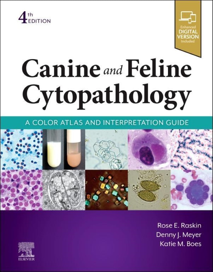
Canine and Feline Cytopathology: A Color Atlas and Interpretation Guide, 4th Edition provides a comprehensive overview of diagnostic cytopathology for companion animals, covering all body systems and fluids. Rapidly resolve diagnostic challenges with this guide to specimen collection and evaluation, featuring more than 2,400 photomicrographs that show cytology of normal structures to contrast and support identification of cytopathology of inflammatory, hyperplastic, and neoplastic lesions. Enhancements to this edition include hundreds of new images with crisper quality and truer colors; new chapters on the pancreas and ear; updated, contemporaneously referenced information for all chapters; expanded listing for neoplastic and infectious disease testing, quality assurance, and reporting; and access to a fully searchable enhanced eBook with new print purchase. Written by seasoned veterinary cytopathologists and award-winning educators Rose Raskin, Denny Meyer, and Katie Boes, with contributions from 20 international experts, this reference offers clear, practical guidelines to sampling procedures, slide preparation, and interpretation leading to diagnoses and/or classification of the cytopathologic findings. Anticipate the expected and expect the unexpected with this atlas, which vividly illustrates the expected cytologic elements associated with the organ system and provides abundant examples of unexpected cytopathologic findings.

Features

Features:

  • Comprehensive coverage of all body systems and body fluidsย emphasizes the application of aspirate biopsy cytopathology for greatest diagnostic impact.
  • Exceptional-quality, full-color photomicrographsย include detailed figure legends.
  • Helpful hints for improving specimen qualityย are provided in discussions of common errors and problems, resulting in more diagnostically effective, cost-effective use of cytopathology.
  • Discussions of clinical findings, differential diagnostic considerations, and the rationaleย are included for the final cytopathological diagnosis.
  • Additional photomicrographs in organ system chaptersย demonstrate the histological or histopathologic corollary of cytopathologic findings.
  • Easy-to-use, well-organized formatย includesย manyย tables, algorithms, boxes, andย Key Pointย callouts for at-a-glance reference.
  • Clear, concise descriptionsย include sampling techniques, slide preparation and examination, and guidelines for interpretation, leading to accurate in-house and commercial laboratory diagnosis.
  • Extensively revisedย organ system-based chapters include focused contemporary references.
Table of Contents

Table of Contents:

Chapter I. Simple Acquistion and Management of Cytology Specimens

Chapter 2. General Categories of Cytologic Interpretation

Chapter 3. Skin and Subcutaneous Tissues

Chapter 4. Hemolymphatic System

Chapter 5. Respiratory Tract

Chapter 6. Body Cavity Effusions

Chapter 7. Oral Cavity, Gastrointestinal Tract, and Associated Structures

Chapter 8. Fecal Cytology

Chapter 9. Pancreas (Exocrine/Endocrine)

Chapter 10. Liver and Gall Bladder

Chapter 11. Urinary Tract

Chapter 12. Microscopic Examination of the Urinary Sediment

Chapter 13. Reproductive System

Chapter 14. Musculoskeletal System

Chapter 15. The Central Nervous System

Chapter 16. Ocular and Otic Sensory Systems

Chapter 17. Endocrine/Neuroendocrine Systems

Chapter 18. Advanced Diagnostic Techniques

Appendix 1. Microscope Equipment and Proper Usage

Appendix 2. Selected Cytologic Staining Protocols

Appendix 3. Cytologically Confusing Structures and Polarizing Materials

Appendix 4. Mitotic Figures and Chromatin Patterns

Appendix 5. Advanced Collection and Preparation Techniques

Appendix 6. Composing Cytologic and Histologic Reports

Appendix 7. List of Selected Specialized Testing Sites

Appendix 8. Quick Reference for Morphologic Features of Infectious and Parasitic Agents

Appendix 9. Quality Assurance and Diagnostic Test Reporting

Get This Book

This Book is Available For Premium Members Only

Become a Premium Now To Get This Book