By Christine C. Lim
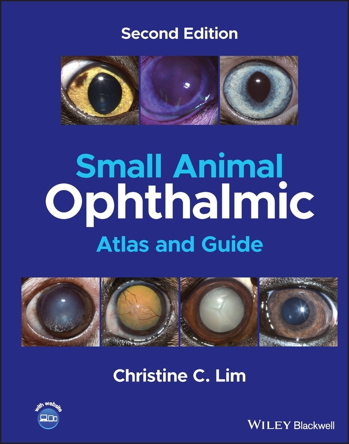
Small Animal Ophthalmic Atlas and Guide 2nd Edition is designed to offer a quick reference to common ocular conditions in dogs and cats, presenting high-quality color photographs to facilitate diagnosis and offering details on each condition to support clinicians in clinical decision making. In addition to updates throughout, the Second Edition includes significantly more images than the previous edition, with updates to images to include more representative examples where possible.
In Small Animal Ophthalmic Atlas and Guide, the image section is organized by area of the eye, making it easy to find and compare images to make a diagnosis, and the disease section is carefully targeted to the most crucial details for developing a management plan. A companion website provides video clips.
With its broad coverage of essential topics and accessible images that help with accurate and fast diagnoses, Small Animal Ophthalmic Atlas and Guide is an essential reference for small animal general practitioners, students, residents, and interns, and can also be used as a reference to show examples to clients.
This Book is Avaiable for Members
Access this book instantly with a Premium membership


![Ettinger’s Textbook of Veterinary Internal Medicine 9th Edition [True PDF+Videos]](https://www.vet-ebooks.com/wp-content/uploads/2024/10/ettingers-textbook-of-veterinary-internal-medicine-9th-edition-100x70.jpg "Ettinger’s Textbook of Veterinary Internal Medicine 9th Edition")


