3D Joint Anatomy In Dogs: Main Joint Pathologies and Surgical Approaches

3D Joint Anatomy In Dogs: Main Joint Pathologies and Surgical Approaches

By Francisco Miguel Sánchez Margallo

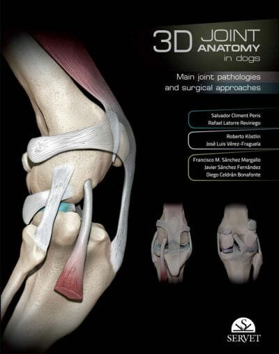
A visual guide with a strongly educational approach covering the main joints in the limbs of the dog. It shows the anatomical elements of each of these joints in three-dimensional diagrams. The views chosen for each case have been selected for a practical purpose, showing the position of the elements involved in the most commonly used surgical approaches. It also describes the key orthopaedic conditions affecting each joint and the most commonly used surgical approaches. It contains a large number of images and illustrations, and a selection of views presented in digital video format.

Table of Contents

1 Stifle joint

Three-dimensional views
Orthopaedic conditions of the stifle joint
Rupture of cruciate ligaments
Clinical signs
Diagnosis
Patellar luxation
Osteochondrosis dissecans (OCD)
Arthritis/osteoarthritis
Diagnostic imaging techniques
Computerised axial tomography
Fluoroscopy (angiography)
Three-dimensional computerised axial tomography
Three-dimensional computerised axial tomography + angiography
Magnetic resonance
Transparent plastinated sections + CAT
Transparent plastinated sections + MRI
Surgical approaches
Approach to the distal portion of the femur and the stifle joint via
lateral incision
Approach to the medial collateral ligament and the caudomedial
region of the stifle joint
Approach to the lateral collateral ligament and the caudolateral
region of the stifle joint
Approach to the proximal portion of the tibia via medial incision

2 Hip joint

Three-dimensional views
Orthopaedic conditions of the hip
Hip dysplasia
Clinical signs
Differential diagnosis
Physical examination
Ortolani test
Treatment
Avascular necrosis of the femoral head
Diagnosis
Differential diagnosis
Treatment
Diagnostic imaging techniques
Transparent plastinated sections + CAT
Transparent plastinated sections + MRI
Surgical approaches
Craniodorsal approach to the hip joint via craniolateral incision
Craniodorsal and caudodorsal approach to the hip joint via osteotomy
of the greater trochanter
Craniodorsal and caudodorsal approach to the hip joint via tenotomy
of the gluteal muscles
Ventral approach to hip joint and pubis

3 Elbow joint

Three-dimensional views
Orthopaedic conditions of the elbow
Fragmented coronoid process (FCP)
Osteochondrosis dissecans (OCD)
Ununited anconeal process (UAP)
Joint incongruity (JI)
Diagnostic imaging techniques
Transparent plastinated sections + CAT
Transparent plastinated sections + MRI
Surgical approaches
Approach to the lateral side of the humeral condyle and epicondyle
Approach to the supracondylar region of the humerus and the caudal
humeroulnar portion of the elbow joint
Approach to the humeroulnar portion of the elbow joint via
osteotomy of the olecranon
Approach to the medial surface of the humeral condyle and medial
coronoid apophysis of the ulna via intermuscular incision

4 Shoulder joint

Three-dimensional views
Orthopaedic conditions of the shoulder
Luxations
Dorsal luxation of the scapula
Luxations of the shoulder joint
Osteochondrosis dissecans of the humeral head
Optimal growth of large breed puppies
Nutrition as a key factor
Diagnosis and treatment
Calcification of the supraspinatus tendon
Treatment
Tenosynovitis of biceps brachii muscle tendon
Treatment
RSI syndrome
Diagnostic imaging techniques
Transparent plastinated sections + CAT
Transparent plastinated sections + MRI
Surgical approaches
Approach to the craniolateral region of the shoulder joint via
tenotomy of the infraspinatus muscle
Approach to the caudolateral region of the shoulder joint
Approach to the craniomedial region of the shoulder joint

5 Carpal and tarsal joints

Three-dimensional views
Orthopaedic conditions of the carpal and tarsal joints
Carpal joint
Carpal luxation and subluxation: hyperextension
Tarsal joint
Tarsal luxation and subluxation
Tibiotarsal luxations
Proximal plantar intertarsal subluxation
Fractures
Medial tarsal abrasion
Osteochondritis dissecans of the talus
Arthrodesis and pan-arthrodesis
Diagnostic imaging techniques
Transparent plastinated sections + CAT
Transparent plastinated sections + MRI
Surgical approaches
Carpal joint
Dorsal approach to the carpal joint
Palmar medial approach to the carpal joint
Tarsal joint
Approach to the lateral malleolus and the talocrural joint
Approach to the medial malleolus and the talocrural joint
Approach to the talocrural joint via osteotomy of the medial
malleolus
Approach to the calcaneus
Approach to the calcaneus and the plantar region of the tarsal bones

6 Bibliography

This Book is For Premium Members Only

Become a Premium Now