By Jean-Marie Denoix
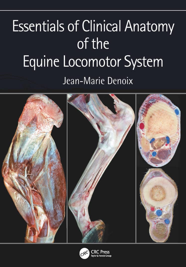
Essentials of Clinical Anatomy of the Equine Locomotor System presents a unique photographic record of dissections showing the topographical anatomy of the locomotor system of the horse. Readers of this book will be able to see the position and relationships of the bones, joints, muscles, nerves and blood vessels that make up each region of the forelimb, vertebral column and hindlimb.
This new atlas is essential for anybody involved in detailed anatomical study, complex lameness evaluation or advanced imaging techniques in horses. It will be a useful guide for veterinary students, and a reference for equine vets in practice.

Recommended Book:
Get This Book
This Book is Avaiable for Members
Access this book instantly with a Premium membership
📄 PDF Format
⚡ Instant Delivery
Already a member? Login


![Ettinger’s Textbook of Veterinary Internal Medicine 9th Edition [True PDF+Videos]](https://www.vet-ebooks.com/wp-content/uploads/2024/10/ettingers-textbook-of-veterinary-internal-medicine-9th-edition-100x70.jpg "Ettinger’s Textbook of Veterinary Internal Medicine 9th Edition")


