back to top

โณ Limited-Time Offer: Get Lifetime Membership With One-Time Payment โ€” Free Updates!

Never Miss A New Book
Sign Up Now
HomeAnimals BooksSmall AnimalsAtlas of Feline Ophthalmology 2nd Edition

Atlas of Feline Ophthalmology 2nd Edition

-

Atlas Of Feline Ophthalmology 2Nd Edition Pdf Download

By Kerry L. Ketring, Mary Belle Glaze

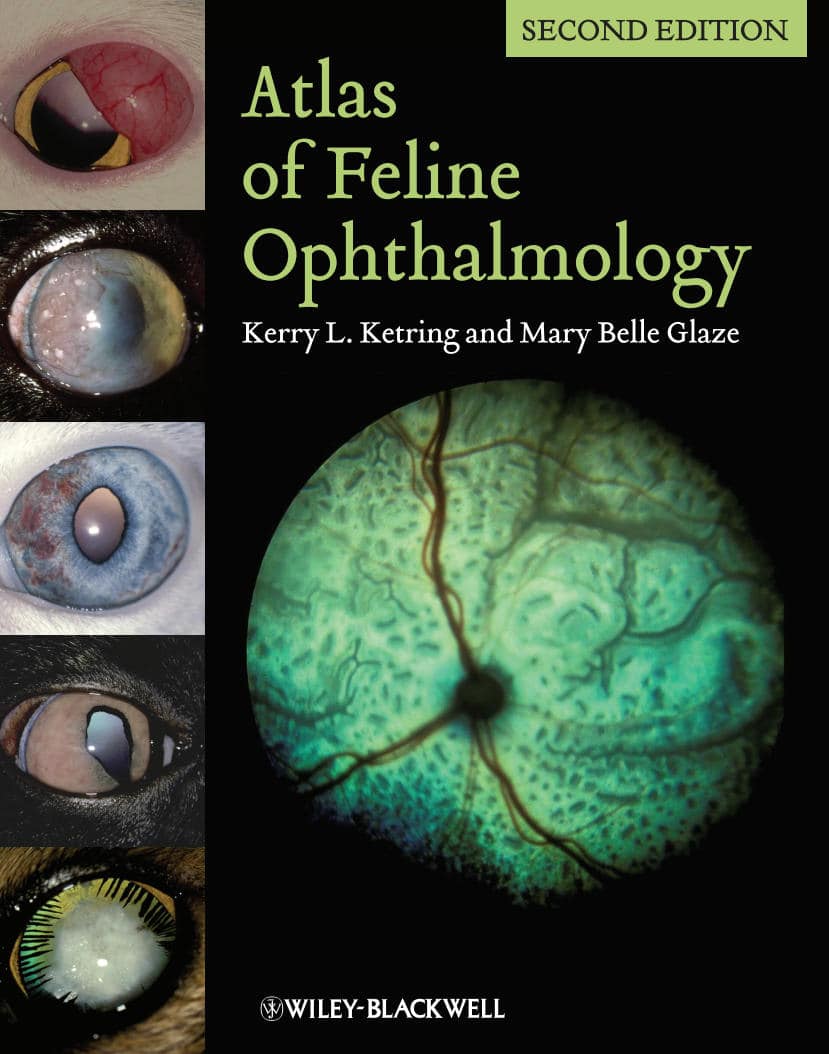
Successful management of eye disease relies on the veterinarianโ€™s ability to identify ocular features and distinguish pathologic changes. Atlas of Feline Ophthalmology 2nd Edition is an invaluable diagnostic reference, providing high-quality color photographs for comparison with a presenting complaint. Presenting 394 photographs illustrating both normal and pathologic ocular conditions, this Second Edition offers a current, complete reference on ocular diseases, adding conditions recognized since publication of the first edition, a broader geographic scope, and many new images with improved quality.

Carefully designed for easy reference, the contents are divided into sections corresponding to specific anatomical structures of the eye. A useful appendix new to this edition groups figures by etiology, making it easy to find every image associated with a specific agent or disease.ย Atlas of Feline Ophthalmology, Second Editionย is a useful tool aiding general practitioners in diagnosing eye disease in cats.

Table of Contents

Table of Contents:

I. Normal Eye

A. Diagrams

B. Normal adnexa/anterior segment

C. Normal fundus

II. Globeโ€“Orbit Relationship

A. Convergent strabismus

B. Enophthalmos

C. Exophthalmos

D. Proptosis

E. Orbitalmucocele

III. Adnexa

A. Eyelid agenesis

B. Entropion

C. Ectropion

D. Distichiasis

E. Blepharitis

F. Apocrine cystadenoma

G. Chalazion

H. Lipogranulomatous conjunctivitis

I. Granuloma/Histoplasmosis

J. Neoplasia

IV. Conjunctiva

A. Dermoid

B. Symblepharon

C. Conjunctivitis

D. Dacryocystitis

E. Neoplasia

V. Nictitating Membrane

A. Nictitans protrusion

B. Eosinophilic conjunctivitis

C. Neoplasia

VI. Cornea

A. Corneal opacities

B. Congenital Endothelial Dysfunction

C. Keratoconus

D. Manx dystrophy

E. Infectious keratitis

F. Ulcerative keratitis

G. Corneal Laceration

H. Eosinophilic keratitis

I. Corneal sequestration

J. Foreign body

K. Staphyloma

L. Neoplasia

VII. Anterior Uvea

A. Dyscorias

B. Persistent pupillary membranes

C. Chediak-Higashi syndrome

D. Iris atrophy

E. Dysautonomia

F. Iris cysts/Iridocilary cysts

G. Anterior uveitis

VIII. Glaucoma

A. Congenital/Goniodysgenesis

B. Inherited/Primary Open Angle Glaucoma (POAG)

C. Feline Aqueous Humor Misdirection Syndrome (FAHMS)

D. Secondary

IX. Lens

A. Senile nuclear sclerosis

B. Cataract

C. Cataract classification by involvement

D. Encephalitozoon cuniculi

E. Lens luxation

X. Vitreous

A. Persistent hyaloid

B. Vitreous hemorrhage

C. Hyalitis

XI. Retina and choroid

A. Congenital

B. Chorioretinitis-Infectious

C. Chorioretinitis-Traumatic

D. Hypertensive retinopathy

E. Retinal detachment

F. Retinal folds

G. Retinopathy

H. Vascular changes

I. Neoplasia

XII. Optic nerve

A. Coloboma

B. Optic disc hypoplasia

C. Optic disc aplasia

D. Optic neuritis

E. Optic nerve atrophy

F. Glaucoma

G. Neoplasia

Get This Book

File Size 29.9 MB
File Format Pdf
Downloadย  link
Useful Links:
All Categories
Join Telegram Channel: Telegram