back to top

Limited-Time Offer: Get Lifetime Membership With One-Time PaymentFree Updates!

Never Miss A New Book
Sign Up Now
HomeAnimals BooksEquineThe Illustrated Horse’s Foot: A comprehensive guide

The Illustrated Horse’s Foot: A comprehensive guide

-

The Illustrated Horse’s Foot: A Comprehensive Guide Pdf Download

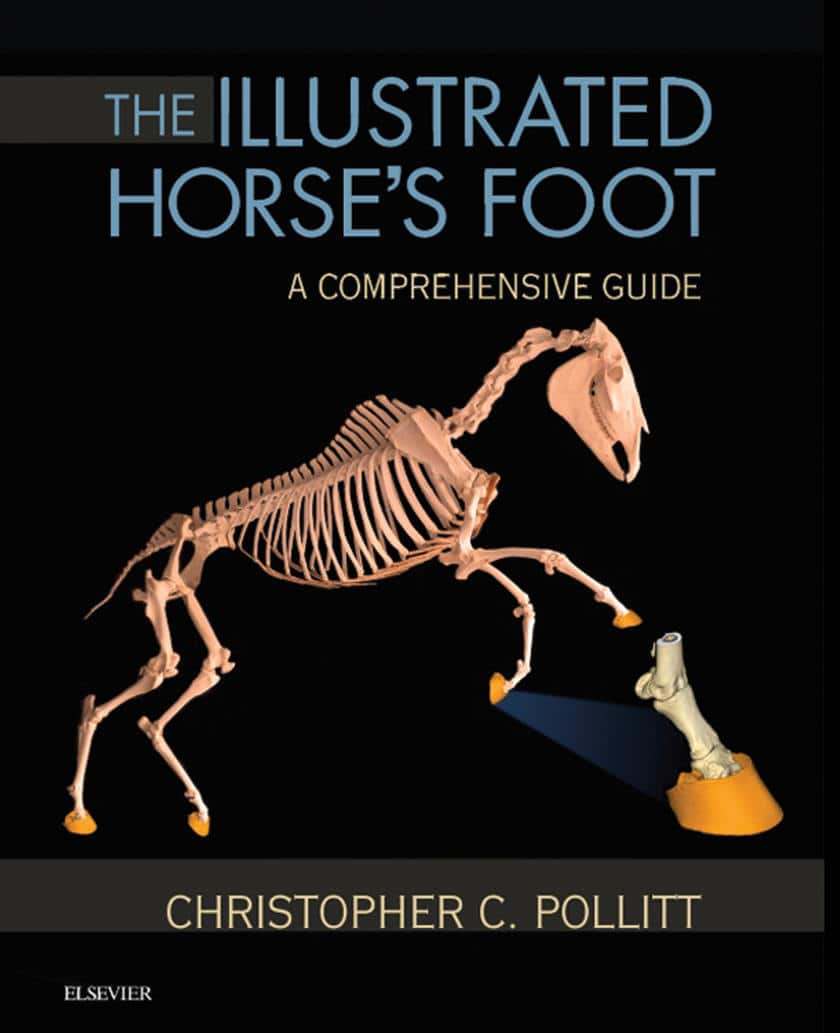
By Christopher C. Pollitt

Achieve optimal results in equine foot care and treatment! The Illustrated Horse’s Foot: A Comprehensive Guide uses clear instructions in an atlas-style format to help you accurately identify, diagnose, and treat foot problems in horses. Full-color clinical photographs show structure and function as well as the principles of correct clinical examination and shoeing, and a companion website has videos depicting equine foot cases. Written by internationally renowned expert Christoher Pollitt, this resource enhances your ability to treat equine conditions ranging from laminitis to foot cracks, infections, trauma, vascular compromise, and arthritis.

Features

Features:

  • Comprehensive coverage addresses a wide range of equine foot conditions.
  • A unique collection of MIMICs provides beautifully detailed anatomical hoof images.
    284 high-quality images show conditions of the equine foot, including many 2-D reconstructions of MRI and CT data.
  • Step-by-step case histories follow equine patients from initial presentation through diagnosis to treatment and outcome.
  • A convenient, templated format provides quick access to clinical signs, diagnosis, treatment, and prognosis.
  • Expert author Chris Pollitt is a pioneer in the use of advanced radiographic, CT, and MRI technology for imaging equine foot and laminitis problems to facilitate accurate diagnosis and effective treatment.
  • A companion website located at pollitthorsesfoot.com located at pollitthorsesfoot.com includes video clips of equine foot cases.
Table of Contents

Table of Contents:

Section 1: Foot Structure and Function

  1. The Hoof

  2. The Hoof Capsule

  3. The Dermis

  4. The Mimics Anatomic Models

  5. Planar Anatomy

  6. The Suspensory Apparatus of the Distal Phalanx

  7. The Circulatory System

  8. Histology

  9. Electron Microscopy

  10. Bones

  11. Joints

  12. Radiography of the Foot

  13. The Palmar Digital Nerve

Section 2: Conditions of the Foot
14. Laminitis
15. Navicular Disease
16. Midline Toe Cracks
17. Seedy Toe (“White Line Disease”)
18. Ossification of the Ungular Cartilages
19. Coronary Band Injury
20. Infected Nail Holes

Index

Get This Book

This Book is Available for Premium Members

Become a Premium Now, Lifetime Deal!