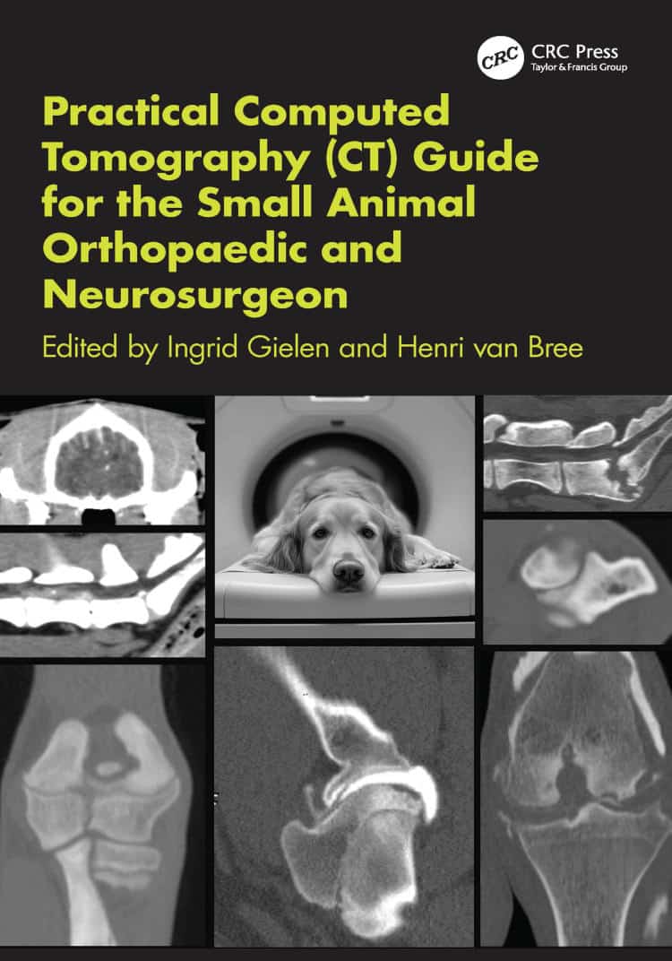
By Ingrid Gielen, Henri van Bree
Practical Computed Tomography (CT) Guide for the Small Animal Orthopaedic and Neurosurgeon addresses the increasing use of veterinary CT in small animal practice, where clinicians are often faced with technical challenges in positioning, equipment handling, and image interpretation.
The book is divided into two parts, beginning with the principles of computed tomography and progressing to body-region–specific CT procedures. With over 200 high-quality images, practical protocols, and clearly structured guidance, it supports accurate CT interpretation and clinical decision-making.
Practical Computed Tomography (CT) Guide for the Small Animal Orthopaedic and Neurosurgeon PDF provides practical support for orthopaedic and neurosurgical imaging, from equipment selection and scanning protocols to diagnosis in small animal patients.
Table of Contents
- Front Matter
- Part I: Principles Of Computed Tomography
- Chapter 1: Introduction And Overview
- Chapter 2: CT Image Characteristics
- Chapter 3: CT Imaging Process And Equipment
- Chapter 4: The Scanning Phase
- Chapter 5: Image Reconstruction
- Chapter 6: Image Display And Viewing
- Chapter 7: Radiation Dose
- Chapter 8: Image Quality And Dose Optimization
- Chapter 9: Patient Preparation
- Chapter 10: Image Interpretation And Diagnosis
- Chapter 11: DICOM Viewing Systems
- Chapter 12: CT System Selection And Purchase
- Chapter 13: CT Procedures And Contrast Applications
- Chapter 14: Cone Beam CT Applications
- Part II: CT Procedures
- Chapter 15: CT Of The Shoulder
- Chapter 16: CT Of The Elbow
- Chapter 17: CT Of The Carpus
- Chapter 18: CT Of The Digits
- Chapter 19: CT Of The Hip And Pelvis
- Chapter 20: CT Of The Stifle
- Chapter 21: CT Of The Tarsus
- Chapter 22: CT Of Long Bones
- Chapter 23: CT Of The Muscles
- Chapter 24: CT Of The Spine
- Chapter 25: CT Of Peripheral Nerves
- Chapter 26: CT Of The Brain
- Chapter 27: Intraoperative And Follow-Up CT
- Appendix
- Back Matter


![Ettinger’s Textbook of Veterinary Internal Medicine 9th Edition [True PDF+Videos]](https://www.vet-ebooks.com/wp-content/uploads/2024/10/ettingers-textbook-of-veterinary-internal-medicine-9th-edition-100x70.jpg "Ettinger’s Textbook of Veterinary Internal Medicine 9th Edition")


