back to top

Limited-Time Offer: Get Lifetime Membership For ONLY $199.99 $149.99 - Deal Will End Soon:

00Hrs 00Mins 00Secs
Sign Up Now
Home Blog Page 76

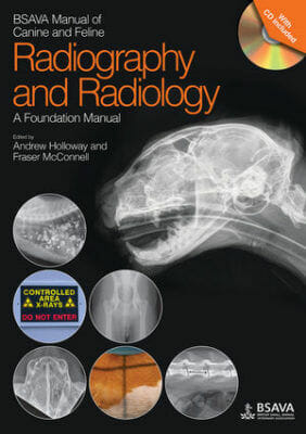
BSAVA Manual of Canine and Feline Radiography and Radiology: A Foundation Manual

BSAVA Manual of Canine and Feline Radiography and Radiology: A Foundation Manual

BSAVA Manual of Canine and Feline Radiography and Radiology: A Foundation Manual 

Bsava Manual Of Canine And Feline Radiography And Radiology: A Foundation Manual

By Andrew Holloway and Fraser McConnell

BSAVA Manual of Canine and Feline Radiography and Radiology: A Foundation Manual PDF. Confident radiographic interpretation presents a considerable challenge and the BSAVA Manual of Canine and Feline Radiography and Radiology provides a comprehensive review of the approach to radiological interpretation, the range of variants and the key fundamental principles and their application to common diseases.

This Foundation Manual replaces the classic BSAVA Manual of Small Animal Diagnostic Imaging as an introduction for veterinary students, nurses and new graduates. The Manual features high quality radiographic reproductions demonstrating normal anatomy and key aspects of interpretation, as well as illustrations showing patient positioning and the practical approach that is the hallmark of the BSAVA Manuals.

Table of Contents

Physics and equipment

Principles of radiography

Principles of radiological interpretation

Contrast radiography

Radiology of the thorax

Radiology of the abdomen

Radiology of the appendicular skeleton

Radiology of the head

Radiology of the vertebral column

Index

Get This Book

This Book is Available For Premium Members Only

Become a Premium Now To Get This Book


Differential Diagnosis in Small Animal Medicine 2nd Edition

Differential Diagnosis in Small Animal Medicine 2nd Edition PDF

Differential Diagnosis in Small Animal Medicine 2nd Edition

Differential Diagnosis In Small Animal Medicine 2Nd Edition Pdf

By Alex Gough and Kathryn F. Murphy

Differential Diagnosis in Small Animal Medicine 2nd Edition PDF is A vital pocket-sized reference tool for busy practitioners and students, saving hours of searching through multiple sources. This Book brings together comprehensive differential diagnosis lists covering a wide range of presenting signs. Differential Diagnosis in Small Animal Medicine 2nd Edition has been fully updated with alphabetised lists for improved navigation. The lists cover the majority of presentations that are encountered in practice, including both common and uncommon conditions.

  • Details differential diagnoses from diverse findings such as history, physical examination, diagnostic imaging, laboratory test results and electrodiagnostic testing
  • Provides guidance on how common conditions are, and how commonly they are the cause of the presenting sign
  • Useful throughout the working day for vets in small animal practice, the information will save hours searching alternative multiple references
  • New co-author Kate Murphy brings her expertise as an ECVIM diplomate

Table of Contents

Introduction 1
Part 1: Historical Signs 
1.1 General, systemic and metabolic historical signs 5
1.1.1 Polyuria/polydipsia 5
1.1.2 Weight loss 7
1.1.3 Weight gain 9
1.1.4 Polyphagia 10
1.1.5 Anorexia/inappetence 11
1.1.6 Failure to grow 13
1.1.7 Syncope/collapse 14
1.1.8 Weakness 18
1.2 Gastrointestinal/abdominal historical signs 22
1.2.1 Ptyalism/salivation/hypersalivation 22
1.2.2 Gagging/retching 24
1.2.3 Dysphagia 26
1.2.4 Regurgitation 27
1.2.5 Vomiting 28
1.2.6 Diarrhoea 34
1.2.7 Melaena 40
1.2.8 Haematemesis 42
1.2.9 Haematochezia 44
1.2.10 Constipation/obstipation 46
1.2.11 Faecal tenesmus/dyschezia 48
1.2.12 Faecal incontinence 49
1.2.13 Flatulence/borborygmus 50
1.3 Cardiorespiratory historical signs 51
1.3.1 Coughing 51
1.3.2 Dyspnoea/tachypnoea 52
1.3.3 Sneezing and nasal discharge 53
1.3.4 Epistaxis 55
1.3.5 Haemoptysis 56
1.3.6 Exercise intolerance 58
1.4 Dermatological historical signs 59
1.4.1 Pruritus 59
1.5 Neurological historical signs 61
1.5.1 Seizures 61
1.5.2 Trembling/shivering 65
1.5.3 Ataxia 67
1.5.4 Paresis/paralysis 76
1.5.5 Coma/stupor 80
1.5.6 Altered behaviour: General changes 82
1.5.7 Altered behaviour: Specific behavioural problems 84
1.5.8 Deafness 85
1.5.9 Multifocal neurological disease 87
1.6 Ocular historical signs 90
1.6.1 Blindness/visual impairment 90
1.6.2 Epiphora/tear overflow 93
1.7 Musculoskeletal historical signs 95
1.7.1 Forelimb lameness 95
1.7.2 Hindlimb lameness 99
1.7.3 Multiple joint/limb lameness 103
1.8 Reproductive historical signs 104
1.8.1 Failure to observe oestrus 104
1.8.2 Irregular seasons 106
1.8.3 Infertility in the female with normal oestrus 107
1.8.4 Male infertility 108
1.8.5 Vaginal/vulval discharge 111
1.8.6 Abortion 111
1.8.7 Dystocia 112
1.8.8 Neonatal mortality 114
1.9 Urological historical signs 115
1.9.1 Pollakiuria/dysuria/stranguria 115
1.9.2 Polyuria/polydipsia 115
1.9.3 Anuria/oliguria 116
1.9.4 Haematuria 117
1.9.5 Urinary incontinence/inappropriate urination 119
Part 2: Physical Signs 121
2.1 General/miscellaneous physical signs 121
2.1.1 Abnormalities of body temperature – hyperthermia 121
2.1.2 Abnormalities of body temperature – hypothermia 127
2.1.3 Enlarged lymph nodes 127
2.1.4 Diffuse pain 130
2.1.5 Peripheral oedema 130
2.1.6 Hypertension 132
2.1.7 Hypotension 133
2.2 Gastrointestinal/abdominal physical signs 135
2.2.1 Oral lesions 135
2.2.2 Abdominal distension 137
2.2.3 Abdominal pain 138
2.2.4 Perianal swelling 141
2.2.5 Jaundice 142
2.2.6 Abnormal liver palpation 144
2.3 Cardiorespiratory physical signs 146
2.3.1 Dyspnoea/tachypnoea 146
2.3.2 Pallor 151
2.3.3 Shock 151
2.3.4 Cyanosis 153
2.3.5 Ascites 155
2.3.6 Abnormal respiratory sounds 155
2.3.7 Abnormal heart sounds 156
2.3.8 Abnormalities in heart rate 160
2.3.9 Jugular distension/hepatojugular reflux 163
2.3.10 Alterations in arterial pulse 163
2.4 Dermatological signs 164
2.4.1 Scaling 164
2.4.2 Pustules and papules (including miliary dermatitis) 166
2.4.3 Nodules 168
2.4.4 Pigmentation disorders (coat or skin) 170
2.4.5 Alopecia 172
2.4.6 Erosive/ulcerative skin disease 174
2.4.7 Otitis externa 176
2.4.8 Pododermatitis 178
2.4.9 Disorders of the claws 180
2.4.10 Anal sac/perianal disease 182
2.5 Neurological signs 183
2.5.1 Abnormal cranial nerve (CN) responses 183
2.5.2 Vestibular disease 186
2.5.3 Horner’s syndrome 189
2.5.4 Hemineglect syndrome (Forebrain dysfunction q.v.) 190
2.5.5 Spinal disorders 190
2.6 Ocular signs 192
2.6.1 Red eye 192
2.6.2 Corneal opacification 197
2.6.3 Corneal ulceration/erosion 198
2.6.4 Lens lesions 200
2.6.5 Retinal lesions 201
2.6.6 Intraocular haemorrhage/hyphaema 203
2.6.7 Abnormal appearance of anterior chamber 204
2.7 Musculoskeletal signs 204
2.7.1 Muscular atrophy or hypertrophy 204
2.7.2 Trismus (‘lockjaw’) 206
2.7.3 Weakness 207
2.8 Urogenital physical signs 207
2.8.1 Kidneys abnormal on palpation 207
2.8.2 Bladder abnormalities 208
2.8.3 Prostate abnormal on palpation 210
2.8.4 Uterus abnormal on palpation 210
2.8.5 Testicular abnormalities 211
2.8.6 Penis abnormalities 211
Part 3: Radiographic and Ultrasonographic Signs 213
3.1 Thoracic radiography 213
3.1.1 Artefactual causes of increased lung opacity 213
3.1.2 Increased bronchial pattern 213
3.1.3 Increased alveolar pattern 214
3.1.4 Increased interstitial pattern 217
3.1.5 Increased vascular pattern 220
3.1.6 Decreased vascular pattern 221
3.1.7 Cardiac diseases that may be associated
with a normal cardiac silhouette 222
3.1.8 Increased size of cardiac silhouette 222
3.1.9 Decreased size of cardiac silhouette 223
3.1.10 Abnormalities of the ribs 224
3.1.11 Abnormalities of the oesophagus 225
3.1.12 Abnormalities of the trachea 228
3.1.13 Pleural effusion 230
3.1.14 Pneumothorax 232
3.1.15 Abnormalities of the diaphragm 233
3.1.16 Mediastinal abnormalities 234
3.2 Abdominal radiography 237
3.2.1 Liver 237
3.2.2 Spleen 239
3.2.3 Stomach 241
3.2.4 Intestines 244
3.2.5 Ureters 251
3.2.6 Bladder 251
3.2.7 Urethra 254
3.2.8 Kidneys 255
3.2.9 Loss of intra-abdominal contrast 258
3.2.10 Prostate 260
3.2.11 Uterus 261
3.2.12 Abdominal masses 261
3.2.13 Abdominal calcification/mineral density 262
3.3 Skeletal radiography 264
3.3.1 Fractures 264
3.3.2 Altered shape of the long bones 264
3.3.3 Dwarfism 265
3.3.4 Delayed ossification/growth
plate closure 266
3.3.5 Increased radiopacity 266
3.3.6 Periosteal reactions 267
3.3.7 Bony masses 267
3.3.8 Osteopenia 268
3.3.9 Osteolysis 270
3.3.10 Mixed osteolytic/osteogenic lesions 271
3.3.11 Joint changes 271
3.4 Radiography of the head and neck 275
3.4.1 Increased radiopacity/bony proliferation
of the maxilla 275
3.4.2 Decreased radiopacity of the maxilla 275
3.4.3 Increased radiopacity/bony proliferation
of the mandible 276
3.4.4 Decreased radiopacity of the mandible 276
3.4.5 Increased radiopacity of the tympanic bulla 276
3.4.6 Decreased radiopacity of the nasal cavity 277
3.4.7 Increased radiopacity of the nasal cavity 277
3.4.8 Increased radiopacity of the frontal sinuses 279
3.4.9 Increased radiopacity of the pharynx 279
3.4.10 Thickening of the soft tissues of the head and neck 280
3.4.11 Decreased radiopacity of the soft tissues
of the head and neck 281
3.4.12 Increased radiopacity of the soft tissues
of the head and neck 281
3.5 Radiography of the spine 282
3.5.1 Normal and congenital variation in vertebral
shape and size 282
3.5.2 Acquired variation in vertebral
shape and size 283
3.5.3 Changes in vertebral radiopacity 285
3.5.4 Abnormalities in the intervertebral space 286
3.5.5 Contrast radiography of the spine
(myelography) 287
3.6 Thoracic ultrasonography 289
3.6.1 Pleural effusion 289
3.6.2 Mediastinal masses 290
3.6.3 Pericardial effusion 290
3.6.4 Altered chamber dimensions 291
3.6.5 Changes in ejection phase indices of left ventricular
performance (fractional shortening,
FS%; ejection fraction, EF) 294
3.7 Abdominal ultrasonography 294
3.7.1 Renal disease 294
3.7.2 Hepatobiliary disease 297
3.7.3 Splenic disease 300
3.7.4 Pancreatic disease 301
3.7.5 Adrenal disease 302
3.7.6 Urinary bladder disease 302
3.7.7 Gastrointestinal disease 304
3.7.8 Ovarian and uterine disease 305
3.7.9 Prostatic disease 306
3.7.10 Ascites 306
3.8 Ultrasonography of other regions 308
3.8.1 Testes 308
3.8.2 Eyes 309
3.8.3 Neck 311
Part 4: Laboratory Findings 313
4.1 Biochemical findings 313
4.1.1 Albumin 313
4.1.2 Alanine transferase 315
4.1.3 Alkaline phosphatase 316
4.1.4 Ammonia 318
4.1.5 Amylase 319
4.1.6 Aspartate aminotransferase 320
4.1.7 Bilirubin 321
4.1.8 Bile acids/dynamic bile acid test 322
4.1.9 C-reactive protein (D) 322
4.1.10 Cholesterol 323
4.1.11 Creatinine 324
4.1.12 Creatine kinase 324
4.1.13 Ferritin 325
4.1.14 Fibrinogen 326
4.1.15 Folate 326
4.1.16 Fructosamine 327
4.1.17 Gamma-glutamyl transferase 327
4.1.18 Gastrin 328
4.1.19 Globulins 329
4.1.20 Glucose 330
4.1.21 Iron 333
4.1.22 Lactate dehydrogenase 333
4.1.23 Lipase 335
4.1.24 Triglycerides 336
4.1.25 Troponin 337
4.1.26 Trypsin-like immunoreactivity 338
4.1.27 Urea 338
4.1.28 Vitamin B12 (cobalamin) 341
4.1.29 Zinc 341
4.2 Haematological findings 342
4.2.1 Regenerative anaemia 342
4.2.2 Poorly/non-regenerative anaemia 345
4.2.3 Polycythaemia 348
4.2.4 Thrombocytopenia 350
4.2.5 Thrombocytosis 353
4.2.6 Neutrophilia 354
4.2.7 Neutropenia 355
4.2.8 Lymphocytosis 357
4.2.9 Lymphopenia 358
4.2.10 Monocytosis 359
4.2.11 Eosinophilia 360
4.2.12 Eosinopenia 361
4.2.13 Mastocytemia 361
4.2.14 Basophilia 362
4.2.15 Increased buccal mucosal bleeding time
(disorders of primary haemostasis) 362
4.2.16 Increased prothrombin time (disorders of
extrinsic and common pathways) 363
4.2.17 Increased partial thromboplastin time or
activated clotting time (disorders of intrinsic
and common pathways) 363
4.2.18 Increased fibrin degradation products 364
4.2.19 Decreased fibrinogen levels 364
4.2.20 Decreased antithrombin III levels 364
4.3 Electrolyte and blood gas findings 365
4.3.1 Total calcium 365
4.3.2 Chloride 367
4.3.3 Magnesium 369
4.3.4 Potassium 371
4.3.5 Phosphate 373
4.3.6 Sodium 375
4.3.7 pH 377
4.3.8 pa02 379
4.3.9 Total C02 381
4.3.10 Bicarbonate 381
4.3.11 Base excess 381
4.4 Urinalysis findings 381
4.4.1 Alterations in specific gravity 381
4.4.2 Abnormalities in urine chemistry 383
4.4.3 Abnormalities in urine sediment 388
4.4.4 Infectious agents 390
4.5 Cytological findings 392
4.5.1 Tracheal/bronchoalveolar lavage 392
4.5.2 Nasal flush cytology 394
4.5.3 Liver cytology 395
4.5.4 Kidney cytology 397
4.5.5 Skin scrapes/hair plucks/tape impressions 398
4.5.6 Cerebrospinal fluid (CSF) analysis 398
4.5.7 Fine-needle aspiration of cutaneous/
subcutaneous masses 400
4.6 Hormones/endocrine testing 401
4.6.1 Thyroxine 401
4.6.2 Parathyroid hormone 403
4.6.3 Cortisol (baseline or post-ACTH stimulation test) 404
4.6.4 Insulin 405
4.6.5 ACTH 405
4.6.6 Vitamin D (1,25-dihydroxycholecalciferol) 405
4.6.7 Testosterone 406
4.6.8 Progesterone 406
4.6.9 Oestradiol 407
4.6.10 Pro-BNP 407
4.7 Faecal analysis findings 408
4.7.1 Faecal blood 408
4.7.2 Faecal parasites 408
4.7.3 Faecal culture 409
4.7.4 Faecal fungal infections 409
4.7.5 Undigested food residues 409
Part 5: Electrodiagnostic Testing 410
5.1 Electrocardiographic findings 410
5.1.1 Alterations in P wave 410
5.1.2 Alterations in QRS complex 411
5.1.3 Alterations in P–R relationship 413
5.1.4 Alterations in S–T segment 414
5.1.5 Alterations in Q–T interval 415
5.1.6 Alterations in T wave 416
5.1.7 Alterations in baseline 416
5.1.8 Rhythm alterations 416
5.1.9 Alterations in rate 420
5.2 Electromyographic findings 422
5.2.1 Spontaneous activity 422
5.2.2 Evoked activity 423
5.3 Nerve conduction velocity findings 423
5.3.1 Decreased velocity 423
5.3.2 Increased velocity 423
Index 424

Get This Book

This Book is Available For Premium Members Only

Become a Premium Now To Get This Book

The Veterinary Book for Dairy Farmers 4th Edition

The Veterinary Book for Dairy Farmers 4th Edition PDF

The Veterinary Book for Dairy Farmers 4th Edition

The Veterinary Book For Dairy Farmers 4Th Edition Pdf

By Roger W. Blowey

The Veterinary Book for Dairy Farmers 4th Edition PDF. The ever-changing world of cattle farming requires farmers to be up-to-date with best-practice procedures and the latest advances in husbandry techniques. Now in its 4th edition, Roger Blowey’s updated version of the acclaimed A Veterinary Book for Dairy Farmers deals with newly emerging problems in cattle farming, as well as covering the necessary knowledge required for maintenance and prosperity. In this practical guide to animal health and husbandry, Blowey highlights developments in established conditions, including lameness, Johnes, BVD, and mastitis. Other updates include the prevention and treatment of emerging diseases, such as Schmallenberg and Blue Tongue, and current issues in farming, such as developments in calf rearing and the increasing incidences of botulism and antimicrobial resistance. This book is a comprehensive and practical text for dairy farmers, stockmen, veterinary academics, and students to navigate this changing field.

Table of Contents

1. A concept of disease, immunity and treatment

2. The young calf

3. The weaned calf

4. Rearing dairy heifers

5. The cow at calving

6. Metabolic disorders

7. Mastitis and conditions of the udder

8. Fertility and its control

9. Lameness and foot trimming

10. Diseases of the skin

11. Notifiable diseases of salmonellosis and zoonoses

12. Minerals, trace elements, vitamins and water

13. Miscellaneous digestive, respiratory and other conditions

14. Routine tasks and dealing with poisons

Get This Book

This Book is Available For Premium Members Only

Become a Premium Now To Get This Book

BSAVA Manual of Canine and Feline Oncology, 3rd Edition

BSAVA Manual of Canine and Feline Oncology, 3rd Edition pdf

Bsava Manual Of Canine And Feline Oncology, 3Rd Edition Pdf

By Jane Dobson and Duncan Lascelles

BSAVA Manual of Canine and Feline Oncology, 3rd Edition PDF. Building on the success of previous editions, the Editors have sought to marry the best of the old with the new. All chapters have been updated, or rewritten, by international experts to encompass the important advances made over the last several years, while keeping the text practical and user-friendly.
The principles of diagnostics and clinical staging and of the main therapeutic modalities are outlined. Individual tumor types in the body systems of dogs and cats are then described, using a common approach to aid information retrieval on etiology and pathogenesis, presentation and clinical signs (including staging/grading of tumors), management, and prognosis. A wealth of new photographs has been included to illustrate the clinical, diagnostic and therapeutic aspects of a range of tumors.

The growing importance of ethical considerations and palliative care are also recognized, and exciting developments and treatment possibilities explored.

Table of Contents

Introduction: cancer in small animals

How to make a diagnosis

Tumour staging

Paraneoplastic syndromes

When to treat animals with cancer

Principles of surgical oncology

Principles of chemotherapy

Principles of radiation therapy

Therapies of the future

Principles of nutrition of the cancer patient

Relief of pain – acute and chronic

Tumours of skin and subcutaneous tissues

Tumours of the skeletal system

Soft tissue sarcomas

Tumours of the mammary glands

Tumours of the urogenital system

Tumours of the respiratory system and thoracic cavity

Tumours of the haemopoietic system

Endocrine tumours

Tumours of the nervous system

Ocular tumours

Index

Get This Book

This Book is Available For Premium Members Only

Become a Premium Now To Get This Book

Georgis’ Parasitology for Veterinarians 11th Edition

Georgis’ Parasitology for Veterinarians 11th Edition PDF Download

Georgis’ Parasitology for Veterinarians 11th Edition

Georgis’ Parasitology For Veterinarians 11Th Edition Pdf Download

By Dwight D. Bowman

Georgis’ Parasitology for Veterinarians 11th Edition provides the most current information on all parasites commonly encountered in veterinary medicine, including minor or rare parasites to assist in the diagnosis of difficult cases. While primarily focused on parasites that infect ruminants, horses, pigs, dogs, and cats, Georgis’ Parasitology for Veterinarians PDF covers organisms that commonly infect laboratory animals and exotic species. More than 600 high-quality, color photographs and illustrations help you learn how to easily identify and treat parasites of every kind.

  • The most comprehensive parasitology content available, written specifically for veterinarians, provides complete information on all parasites commonly encountered in veterinary medicine, as well as information about minor or rare parasites.
  • High-quality color photographs and illustrations make the process of identifying and treating parasites more accurate and efficient.
Table of Contents

1.Introduction
2. Arthropods
3. Protozoans
4. Helminths
5. Vector-Borne Diseases
6. Antiparasitic Drugs
7. Diagnostic Parasitology
8. Histopathologic Diagnosis
9. Vaccinations

Get This Book

This Book is Available For Premium Members Only

Become a Premium Now To Get This Book

Complications in Small Animal Surgery

Complications in Small Animal Surgery PDF

Complications in Small Animal Surgery

Complications In Small Animal Surgery Pdf

By Dominique Griffon, Annick Hamaide

Complications in Small Animal Surgery PDF provides a complete reference to diagnosing, managing, and treating surgical complications, with information following a standardized format for ease of use.

•     Presents comprehensive information on diagnosing, managing, and preventing surgical complications using an accessible format
•     Offers a  well-defined, thoroughly illustrated format to maximize practical value, with algorithms, tables, practical tips, and many images throughout
•     Covers common and uncommon complications in all body systems
•     Serves as a reference to recent literature relevant to each complication
•     Includes access to a companion website with videos, figures from the book available for download into

Table of Contents

List of Contributors, ix

Preface and Acknowledgements, xiii

About the companion website, xv

Section 1: Surgical Infections, 1

1 Surgical Wound Infections, 3
Outi Laitinen-Vapaavuori

2 Sepsis, 8
Galina Hayes and Brigitte A. Brisson

3 Pyothorax, 15
Jackie L. Demetriou

4 Peritonitis, 20
Marjan Doom and Hilde de Rooster

5 Osteomyelitis, 28
Dominique Griffon

6 Septic Arthritis, 34
John Innes

7 Methicillin-Resistant Staphylococcus Infections, 39
Scott Weese

8 Hospital-Associated Infections, 45
Maureen Anderson and Scott Weese

Section 2: General Postoperative Complications, 55

9 Dehiscence, 57
Stéphanie Claeys

10 Suture Reactions, 64
Outi Laitinen-Vapaavuori

11 Hyperthermia, 66
Kris Gommeren

12 Hemorrhage, 72
Jane Ladlow

13 Anorexia, 79
Angela Witzel

14 Management of Surgical Pain, 84
Lyon Lee and Reza Seddighi

15 Complications Associated with, Nonsteroidal Antiinflammatory Drugs, 97
Reza Seddighi and Lyon Lee

16 Complications Associated with External Coaptation, 110
Dominique Griffon

17 Complications Associated with Rehabilitation Modalities, 118
Debra Canapp, Lisa M. Fair, and Sherman Canapp

18 Degenerative Joint Disease, 126
Guillaume Ragetly and Dominique Griffon

Section 3: Surgery of the Head and Neck, 135

19 Dorsal Rhinotomy, 137
Gertter Haar

20 Sublingual and Mandibular Sialadenectomy, 144
Cheryl Hedlund

21 Incisional Drainage of Aural Hematomas, 150
Cheryl Hedlund

22 Ear Canal Surgery, 155
Gertter Haar

23 Ventral Bulla Osteotomy, 164
Cheryl Hedlund

24 Surgical Management of Brachycephalic Airway Obstructive Syndrome, 171
Cyril Poncet

25 Surgical Management of Laryngeal Paralysis, 178
Gertter Haar

26 Thyroidectomy, 185
Chad Schmiedt

27 Parathyroidectomy, 193
Chad Schmiedt

28 Tracheal Surgery, 198
Barbara Kirby

29 Tracheal Stenting, 206
Laura Cuddy

Section 4: Dental and Oral Surgery, 215

30 Repair of Mandibular and Maxillary Fractures, 217
Christian Bolliger

31 Partial Mandibulectomy, 229
Julius Liptak

32 Partial Maxillectomy, 237
Julius Liptak

33 Lingual Surgery, 245
Julius Liptak

34 Tooth Extractions, 249
Anson J. Tsugawa

35 Tonsillectomy, 263
Jonathan Bray

36 Repair of Congenital Defects of the Hard and Soft Palates, 265
Jonathan Bray

Section 5: Thoracic Surgery, 269

37 General Complications, 271
Hervé Brissot

38 Thoracostomy and Th oracocentesis, 284
Alison Moores

39 Complications After Intercostal and Sternal Thoracotomy, 294
Alison Moores

40 Thoracoscopy, 300
Eric Monnet

41 Bronchial and Pulmonary Surgery, 305
Hervé Brissot

42 Chylothorax, 313
Carolyn A. Burton

43 Esophagoscopy and Esophageal Surgery, 319
Robert J. Hardie

44 Aortic Arch Anomalies, 323
Robert J. Hardie

45 Thymectomy, 328
Rachel Burrow

46 Pericardial Surgery, 335
Daniel J. Brockman

47 Patent Ductus Arteriosus, 338
Daniel J. Brockman

48 Cardiac Pacemaker Implantation, 343
Rachel Burrow and Sonja Fonfara

Section 6: General Abdominal Surgery, 353

49 Celiotomy, 355
Hilde de Rooster

50 Laparoscopy, 362
Gilles Dupré and Valentina Fiorbianco

51 Hiatal Herniorrhaphy, 370
Stephen J. Baines

52 Diaphragmatic Herniorrhaphy, 375
Stephen J. Baines

53 Peritoneal Pericardial Herniorrhaphy, 383
Laura J. Owen

54 Perineal Herniorrhaphy, 388
Lynne A. Snow

55 Adrenalectomy, 395
Brigitte Degasperi and Gilles Dupré

56 Splenectomy, 401
Paolo Buracco and Federico Massari

Section 7: Gastrointestinal Surgery, 411

57 Gastric Dilatation and Volvulus and Gastropexies, 413
Zoë Halfacree

58 Gastric Outflow Procedures, 424
Jeffrey J. Runge

59 Small Intestine Surgery, 429
Jean-Philippe Billet

60 Large Intestine Surgery, 435
Jean-Philippe Billet

61 Hepatic and Biliary Tract Surgery, 441
Anne Kummeling

62 Pancreatic Surgery, 446
Heidi Phillips

63 Portosystemic, 461
Barbara Kirby

Section 8: Surgery of the Urinary Tract, 467

64 General Complications, 469
Anne Kummeling

65 Complications Specific to Lithotripsy, 473
Joseph W. Bartges, Amanda Callens, and India Lane

66 Renal Transplantation, 481
Andrew Kyles

67 Ureteral Surgery, 486
Stéphanie Claeys and Annick Hamaide

68 Surgery of the Urinary Bladder, 492
Annick Hamaide

69 Urethrostomy in Male Dogs, 497
Joshua Milgram

70 Feline Perineal Urethrostomy, 500
Joshua Milgram

Section 9: Surgery of the Reproductive Tract, 505

71 Ovariectomy and Ovariohysterectomy, 507
Annick Hamaide

72 Pyometra, 517
Kathryn Pratschke

73 Cesarean Section, 522
Bart Van Goethem

74 Orchiectomy, 528
Bart Van Goethem

75 Prostatic Cyst and Abscess Drainage, 534
Henry L’Eplattenier

76 Prostatectomy, 539
Henry L’Eplattenier

Section 10: Plastic and Reconstructive Surgery, 543

77 Open Wounds, 545
Davina Anderson

78 Reconstructive Flaps, 554
Stéphanie Claeys

79 Skin Grafting, 561
Stéphanie Claeys

80 Anal Sac Excision, 569
Lynne A. Snow

81 Feline Onychectomy, 573
Ameet Singh and Brigitte A. Brisson

Section 11: Neurologic Surgery, 577

82 General Complications, 579
Belinda Comito

83 Myelography, 584
Belinda Comito

84 Ventral Slot and Fenestration, 590
Wanda Gordon-Evans

85 Dorsal Laminectomy, 596
Wanda Gordon-Evans

86 Hemilaminectomy, 602
Wanda Gordon-Evans

87 Ventral Cervical Fusion, 606
Wanda Gordon-Evans

88 Vertebral Fracture Repair, 610
Wanda Gordon-Evans

89 Craniotomy and Craniectomy, 615
Belinda Comito

90 Intracranial Shunt Placement, 620
Belinda Comito

Section 12: Management of Fractures and Osteotomies: Perioperative Complications, 627

91 Inadequate Fracture Reduction (Malreduction), 629
Ann Johnson

92 Improper Placement of Implants, 633
Ann Johnson

93 Limb Shortening, 637
Ann Johnson

94 Implant Failure, 641
Ann Johnson

95 Premature Physeal Closure After Fracture, 649
Ann Johnson

96 Avascular Necrosis, 655
Dominique Griffon

97 Bone Resorption, 658
Dominique Griffon

98 Delayed Union, 665
Ann Johnson

99 Nonunion, 669
Ann Johnson

100 Malunion, 673
Andrew Worth

101 Loss of Function in Performance Dogs, 680
Lauren Pugliese and Jon Dee

102 Carpal Contracture, 686
Alessandro Piras and Tomas Guerrero

103 Quadriceps Contracture, 692
Darryl Millis

104 Fracture-Associated Sarcoma, 697
G. Elizabeth Pluhar

105 Thermal Pain, 702
G. Elizabeth Pluhar

106 Complications in External Skeletal Fixation, 704
Gian Luca Rovesti

107 Complications Specific to Locking Plates, 714
Randy Boudrieau

Section 13: Salvage Orthopedic Procedures, 727

108 Arthrodesis, 729
Dominique Griffon

109 Amputation, 735
Maria Fahie

110 Limb Spare Procedures, 743
Jennifer Covey and Nicholas Bacon

Section 14: Surgery of the Coxofemoral Joint, 751

111 Femoral Head and Neck Excision, 753
Wing Tip Wong

112 Complications of Double and Triple Pelvic Osteotomies, 759
Aldo Vezzoni

113 Total Hip Replacement, 778
William Liska and Jonathan Dyce

114 Complications of Total Hip Replacement with the Zurich Cementless System, 834
Aldo Vezzoni

115 Open Reduction of Coxofemoral Luxations, 845
Mark Rochat

Section 15: Surgery of the Stifle, 857

116 Complications Associated with Open versus Arthroscopic Examination of the Stifle, 859
Antonio Pozzi and Stanley Kim

117 Complications associated with cranial cruciate ligament repair, 867
Dominique Griffon

118 Complications Associated with the Treatment of Patellar Luxation, 889
Abimbola Oshin

119 Complications Associated with Autogenous Osteochondral Repair, 897
Noel Fitzpatrick

Section 16: Surgery of the Scapulohumeral Joint, 903

120 Shoulder Dislocation and Instability, 905
Brian Beale

Section 17: Surgery of the Elbow, 917

121 Reduction of Elbow Luxations, 919
Peter Böttcher

122 Ulnar Osteotomy and Ostectomy, 924
Dominique Griffon

123 Complications of Sliding Humeral Osteotomy, 930
Noel Fitzpatrick

Index, 937
Get This Book

This Book is Available For Premium Members Only

Become a Premium Now To Get This Book


Complications in Canine Cranial Cruciate Ligament Surgery

Complications in Canine Cranial Cruciate Ligament Surgery PDF

Complications in Canine Cranial Cruciate Ligament Surgery

Complications In Canine Cranial Cruciate Ligament Surgery Pdf

By David L. Dycus and Ron Ben-Amotz

Filling a gap in the current literature, Complications in Canine Cranial Cruciate Ligament Surgery PDF provides revision strategies for correcting the complications associated with surgical repair techniques for cranial cruciate ligament rupture, one of the most common causes of a hind limb lameness in dogs. Presenting step-by-step instructions for numerous surgical correction techniques, this practical guide covers articular, extra-articular and osteotomy repair techniques as well as non-surgical management, physical rehabilitation, clinical decision making, and more.

Complications in Canine Cranial Cruciate Ligament Surgery PDF begins with an overview of cranial cruciate ligament tear, diagnosis, and treatment goals, followed by a discussion of methods for minimizing surgical site infection and complications. Subsequent chapters describe the potential complications of a particular technique and explain how to identify, evaluate, and correct the complication. Throughout the book, hundreds of high-quality clinical photographs show the appearance of complications and demonstrate each step of the corrective procedure. This authoritative guide:

  • Provides step-by-step techniques for surgical corrections of common complications
  • Emphasizes surgical decision making and specific strategies for surgical correction
  • Contains revision strategies for identification of intra-operative complications
  • Covers evaluation and identification of post-operative complications
  • Features more than 400 photographs and clinical images

Part of the state-of-the-art Advances in Veterinary Surgery series, Complications in Canine Cranial Cruciate Ligament Surgery is an invaluable resource for surgical residents, veterinary surgeons, and general practice veterinarians alike.

Table of Contents

List of Contributors vii
Preface ix
Foreword xi
Acknowledgments xiii
Disclosures xv
I Introduction 1
1 Pathology, Diagnosis, and Treatment
Goals of Cranial Cruciate Ligament
Rupture
and Defining Complications 3
David L. Dycus, Jeffery Biskup, Michael G.
Conzemius, and Ron Ben‐Amotz
2 Surgeon and Patient Preparation to
Minimize
Surgical Site Complications
and Infection
Surveillance Programs 15
Katie L. Hoddinott, J. Scott Weese,
and Ameet Singh
3 Identification, Addressing, and
Following
Up on Surgical Site Infection
After Cranial Cruciate Ligament
Stabilization
33
Katie L. Hoddinott, J. Scott Weese,
and Ameet Singh
II Intraarticular Repair
Techniques 43
4 Complications Associated with
Intraarticular Repair Techniques 45
Jeffery Biskup and Michael G. Conzemius
5 Complications Associated
with Proximal Tibial
Epiphysiodesis 61
Aldo Vezzoni, Joshua S. Zuckerman,
and Ron Ben‐Amotz
III Extraarticular Repair
Techniques 85
6 Extracapsular Stabilization Using
Synthetic Material 87
Ian G. Holsworth
7 Complications Associated with
Extracapsular
Stabilization Using
Monofilament Material 109
R. Randy Basinger
8 Complications Associated with
Extracapsular
Stabilization using
Multifilament
Material 121
Matt Corse and Ian G. Holsworth
IV Osteotomy Repair Techniques 145
9 Complications Associated with
Cranial Closing Wedge Osteotomy 147
Bill Oxley
10 Complications Associated with Tibial
Plateau
Leveling Osteotomy 163
Ron Ben‐Amotz and Joshua S. Zuckerman
11 Complications Associated with
CORA‐Based Leveling Osteotomy 189
David L. Dycus and Don Hulse

12 Complications Associated with Tibial
Tuberosity Advancement 199
Matthew D. Barnhart
13 Complications Associated with the
Modified Maquet Technique 217
Marc H. Balligand
V Nonsurgical Management and
Physical Rehabilitation 235
14 Complications Associated with Stifle
Orthotics 237
Bryan T. Torres and Steven C. Budsberg
15 Complications from Cranial Cruciate
Ligament Surgery: Rehabilitation
Considerations
247
Matthew W. Brunke, David Levine, David L.
Dycus, and Denis J. Marcellin‐Little
VI Adjunctive Information 261
16 Complications Associated with Feline
Cranial Cruciate Ligament Techniques 263
Sorrel J. Langley-Hobbs and
Eva Schnabl‐Feichter
17 Complications Associated with
Arthroscopic Evaluation of the Stifle
and Decision Making 287
Brian S. Beale, Stephen C. Jones,
and Selena Tinga
18 Complications Associated with the
Meniscus and Decision Making 307
Stephen C. Jones, Selena Tinga, and
Brian S. Beale
Index

Get This Book

This Book is Available For Premium Members Only

Become a Premium Now To Get This Book

Small Animal Veterinary Psychiatry

Small Animal Veterinary Psychiatry PDF

Small Animal Veterinary Psychiatry

Small Animal Veterinary Psychiatry Pdf

By Sagi Denenberg

Small Animal Veterinary Psychiatry PDF. Problem behaviours are often the result of how an animal thinks and feels, genetics and environmental influences. Steering away from just description diagnoses and focusing instead on emotional and cognitive causes, this book provides a practical approach to diagnosing, treating, and managing behaviour pathologies in dogs and cats.

Beginning by addressing cases in the first opinion practice, Small Animal Veterinary Psychiatry PDF then considers physical disorders that may lead to or exacerbate abnormal behavior. From there, the focus shifts to mental and emotional health, from an assessment of normal behavior and giving juveniles an optimal start in life, to diagnosing mental and emotional disorders, addressing emotions such as anxiety and frustration, and how to manage these issues – by modifying behavior, managing the animal’s environment, training, and, when necessary, the use of medications. The second half of the book then addresses owner concerns, including management problems, aggression, affective disorder, elimination disorder, abnormal and repetitive behaviours and ageing-related problems.

With an emphasis on helping first line veterinarians identify common presentations and offer help to owners, this book:
– Addresses both normal and abnormal behaviour in cats and dogs from an emotion and cognition perspective;
– Provides behaviour modification protocols, and drug doses and indications;
– Includes handouts to be used both within the practice and with clients to help the veterinary surgeon manage the case.

Written by international experts, the book translates their insights and experience into approaches taken in behavioural medicine. Also including the most up-to-date drugs, it is an important resource for both small animal veterinarians and students of veterinary medicine or animal behaviour.

Table of Contents

Table of contents

1: Addressing Mental and Emotional Health in the Veterinary Practice.

2: Ruling Out Physical Disorders Leading to Behavioural Changes.

3: Addressing Pain in Veterinary Psychiatry.

4: Normal Behaviour – Cats.

5: Normal Behaviour – Dogs.

6: Raising Mentally and Emotionally Healthy Pets.

7: Diagnosis.

8: Learning Principles and Behaviour Modification.

9: Psychopharmacology.

10: Problem Behaviours and Management.

11: Aggression – Cats.

12: Aggression – Dogs.

13: Affective Disorders in Cats and Dogs.

14: Elimination Problems in Cats and Dogs.

15: Abnormal and Repetitive Behaviours in Cats and Dogs.

16: Ageing-related Problems in Cats and Dogs.

Get This Book

This Book is Available For Premium Members Only

Become a Premium Now To Get This Book

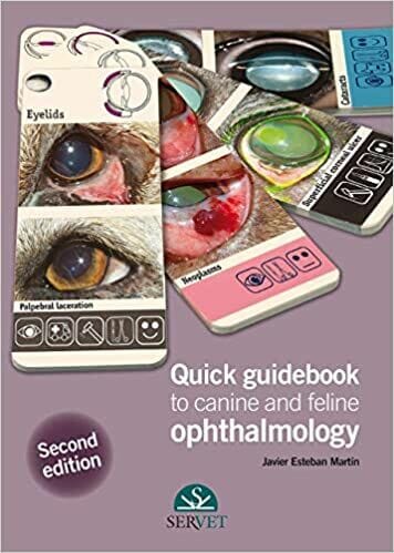
Quick Guidebook to Canine and Feline Ophthalmology 2nd Edition

Quick Guidebook to Canine and Feline Ophthalmology 2nd Edition

Quick Guidebook to Canine and Feline Ophthalmology 2nd Edition

Quick Guidebook To Canine And Feline Ophthalmology 2Nd Edition

By Javier Esteban Martín

Quick Guidebook to Canine and Feline Ophthalmology 2nd Edition PDF provide both the veterinary surgeon and the student with a quick guide that will allow them to easily recognise the ophthalmological conditions most commonly seen in daily practice. All the images shown in this guide correspond to real-lifecases and are accompanied by brief texts that facilitate their understanding and turn this book into an excellent reference aid.


Table of Contents

Organisation of this guidebook

How to use this guidebook

Key for the Quick guidebook to canine and feline ophthalmology

1 Anatomical structures

Diagram of the eye and its adnexa

Lacrimal apparatus diagram

2 Eyelids

Palpebral agenesis

Palpebral dermoid

Entropion in a Shar Pei

Entropion in a Chow Chow

Entropion (surgical techniques)

Medial entropion

Lateral entropion

Cicatricial entropion

Entropion due to lack of support

Ectropion

Entropion/ectropion

Entropion/ectropion. Diamond eye

Euryblepharon

Palpebral laceration

Fungal blepharitis

Bacterial blepharitis

Parasitic blepharitis

Blepharitis (hypersensitivity)

Immune blepharitis

Palpebral neoplasms

Distichiasis

Districhiasis

Ectopic cilia

Trichiasis

Trichomegaly

Palpebral ptosis

Lagophthalmos

Chalazion

Hordeolum

Lentigo

Foreign bodies

3 Nictitating membrane

Prolapse of the nictitating membrane gland

Cartilage eversion

Plasmoma

Follicular conjunctivitis

Granulomas due to Leishmania

Lacerations/tears

Protrusion of the nictitating membrane

Neoplasms

Foreign bodies

Depigmentation of the free edge of the nictitating membrane

4 Conjunctiva

Conjunctival dermoid

Conjunctival cysts

Conjunctivitis

Keratoconjunctivitis sicca

Symblepharon

Drug plaques

Mucinosis

Haemorrhages

Chemosis

Wounds

Neoplasms

5 Cornea and sclera

Superficial corneal ulcer

Refractory corneal ulcer

Stromal ulcer

Deep corneal ulcer

Descemetocoele

Corneal perforation

Melting corneal ulcers

Chronic superficial keratitis

Keratoconjunctivitis sicca

Pigmentary keratitis

Feline eosinophilic keratitis

Corneal dystrophy

Corneal degeneration

Corneal oedema

Neoplasms

Foreign bodies

Feline corneal sequestrum

Dermoid cyst

Corneal abscess

Episcleritis

6 Uveal tract

Iris hypoplasia

Iris heterochromia

Persistent pupillary membrane

Uveitis

Iris cysts

Neoplasms

Iris atrophy

Melanosis

Hyphaema

7 The lens

Microphakia

Coloboma

Nuclear sclerosis

Cataracts

Lens subluxation

Lens luxation

8 Ocular fundus

Normal ocular fundus

Retinal detachment

Retinal haemorrhage

Retinal dysplasia

Optic nerve hypoplasia

Optic neuritis

Retinal degeneration

9 Globe and orbit

Enophthalmos

Exophthalmos

Atrophy of the globe

Buphthalmos

Retrobulbar abscess

Orbital cellulitis

Orbital neoplasms

Foreign bodies

Proptosis

10 Lacrimal apparatus

Epiphora

Keratoconjunctivitis sicca

Imperforate lacrimal punctum

Hypoplasia

Obstruction

Dacryocystitis

11 Glaucoma

Diagnosis

Classification

Clinical signs

Differential diagnosis

Treatment

Sequelae

Bibliography

Alphabetical index

Get This Book

This Book is Available For Premium Members Only

Become a Premium Now To Get This Book

Minding Dogs: Humans, Canine Companions, and a New Philosophy of Cognitive Science

Minding Dogs: Humans, Canine Companions, and a New Philosophy of Cognitive Science PDF By Michele Merritt

Minding Dogs: Humans, Canine Companions, And A New Philosophy Of Cognitive Science Pdf By Michele Merritt

By Michele Merritt

Minding Dogs: Humans, Canine Companions, and a New Philosophy of Cognitive Science PDF. While the past decade has seen a surge of research regarding canine cognition, this newfound interest has not caught the attention of many philosophers. Studies pertaining to dog minds have been pouring out of canine cognition labs all over the world, but they remain relatively ensconced within the scientific, sociological, and anthropological communities, and very little philosophical thought on dog cognition exists.

Philosophers certainly have not shied away from theorizing about the nature of nonhuman animal cognition generally. Theories range from Cartesian disavowal of all nonhuman intelligence to arguments that even fish have complex minds and therefore humans should not eat them. Serious philosophical considerations about dogs and their relationship to humans, however, remain incredibly rare. Even less common, if not entirely nonexistent, is a
critical examination of the question “What are dogs thinking?” and what asking and attempting to answer this question reveals, not so much about dogs, but about us.

With Minding Dogs Michele Merritt attempts to fill two significant gaps in the philosophy of animal cognition. First, she adds to the growing discussion on canine cognition, which has been overlooked until recently and requires more consideration. Second, she takes seriously our dynamic collaborations with our canine friends as crucial to understanding both their minds and our own.

Table of Contents

INTRODUCTION

CHAPTER 1 Canine Minds: Historical Precedents and Current Curiosity

CHAPTER 2 Thinking-with Dogs and Dismantling Standard Cognitive Science

CHAPTER 3 Canine Mindreading and Interspecies Social Cognition

CHAPTER 4 Thinking-in-Playing: Social Cognition beyond Mindreading

CHAPTER 5 Dynamic Duos: Making-with, Thinking-with, and Enacting Interspecies Collaborations

POSTSCRIPT Six-Million-Dollar Dogs and the Future of Companion Species Studies

NOTES

WORKS CITED

INDEX

Get This Book

This Book is Available For Premium Members Only

Become a Premium Now To Get This Book